7NKU

diazaborine bound Drg1(AFG2)


Experimental Data Snapshot

  • Method: ELECTRON MICROSCOPY
  • Resolution: 3.40 Å
  • Aggregation State: PARTICLE 
  • Reconstruction Method: SINGLE PARTICLE 

wwPDB Validation   3D Report Full Report


This is version 1.0 of the entry. See complete history


Literature

Structural basis for inhibition of the AAA-ATPase Drg1 by diazaborine.

Prattes, M.Grishkovskaya, I.Hodirnau, V.V.Rossler, I.Klein, I.Hetzmannseder, C.Zisser, G.Gruber, C.C.Gruber, K.Haselbach, D.Bergler, H.

(2021) Nat Commun 12: 3483-3483

  • DOI: https://doi.org/10.1038/s41467-021-23854-x
  • Primary Citation of Related Structures:  
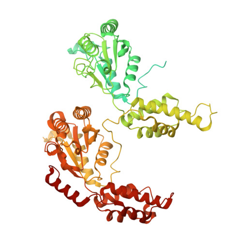
    7NKU

  • PubMed Abstract: 

    The hexameric AAA-ATPase Drg1 is a key factor in eukaryotic ribosome biogenesis and initiates cytoplasmic maturation of the large ribosomal subunit by releasing the shuttling maturation factor Rlp24. Drg1 monomers contain two AAA-domains (D1 and D2) that act in a concerted manner. Rlp24 release is inhibited by the drug diazaborine which blocks ATP hydrolysis in D2. The mode of inhibition was unknown. Here we show the first cryo-EM structure of Drg1 revealing the inhibitory mechanism. Diazaborine forms a covalent bond to the 2'-OH of the nucleotide in D2, explaining its specificity for this site. As a consequence, the D2 domain is locked in a rigid, inactive state, stalling the whole Drg1 hexamer. Resistance mechanisms identified include abolished drug binding and altered positioning of the nucleotide. Our results suggest nucleotide-modifying compounds as potential novel inhibitors for AAA-ATPases.


  • Organizational Affiliation

    Institute of Molecular Biosciences, University of Graz, Graz, Austria.


Macromolecules
Find similar proteins by:  (by identity cutoff)  |  3D Structure
Entity ID: 1
MoleculeChains Sequence LengthOrganismDetailsImage
ATPase family gene 2 protein
A, B, C, D, E
A, B, C, D, E, F
780Saccharomyces cerevisiae S288CMutation(s): 0 
Gene Names: AFG2DRG1YLR397CL8084.16
EC: 3.6.4.10
UniProt
Find proteins for P32794 (Saccharomyces cerevisiae (strain ATCC 204508 / S288c))
Explore P32794 
Go to UniProtKB:  P32794
Entity Groups  
Sequence Clusters30% Identity50% Identity70% Identity90% Identity95% Identity100% Identity
UniProt GroupP32794
Sequence Annotations
Expand
  • Reference Sequence
Small Molecules
Ligands 2 Unique
IDChains Name / Formula / InChI Key2D Diagram3D Interactions
AGS
Query on AGS

Download Ideal Coordinates CCD File 
G [auth A]
H [auth A]
K [auth B]
L [auth B]
N [auth C]
G [auth A],
H [auth A],
K [auth B],
L [auth B],
N [auth C],
O [auth C],
Q [auth D],
R [auth D],
T [auth E],
U [auth E],
W [auth F],
X [auth F]
PHOSPHOTHIOPHOSPHORIC ACID-ADENYLATE ESTER
C10 H16 N5 O12 P3 S
NLTUCYMLOPLUHL-KQYNXXCUSA-N
TDB (Subject of Investigation/LOI)
Query on TDB

Download Ideal Coordinates CCD File 
I [auth A]
J [auth A]
M [auth C]
P [auth D]
S [auth D]
I [auth A],
J [auth A],
M [auth C],
P [auth D],
S [auth D],
V [auth F]
6-METHYL-2(PROPANE-1-SULFONYL)-2H-THIENO[3,2-D][1,2,3]DIAZABORININ-1-OL
C9 H13 B N2 O3 S2
TVXLILKNSPCVRB-UHFFFAOYSA-N
Experimental Data & Validation

Experimental Data

  • Method: ELECTRON MICROSCOPY
  • Resolution: 3.40 Å
  • Aggregation State: PARTICLE 
  • Reconstruction Method: SINGLE PARTICLE 
EM Software:
TaskSoftware PackageVersion
RECONSTRUCTIONcryoSPARC
MODEL REFINEMENTRosetta
MODEL REFINEMENTPHENIX

Structure Validation

View Full Validation Report


Entry History 

Deposition Data

Revision History  (Full details and data files)

  • Version 1.0: 2021-06-23
    Type: Initial release