7NKG

Methyl-coenzyme M reductase from Methermicoccus shengliensis at 1.6-A resolution


Experimental Data Snapshot

  • Method: X-RAY DIFFRACTION
  • Resolution: 1.60 Å
  • R-Value Free: 0.190 
  • R-Value Work: 0.172 
  • R-Value Observed: 0.173 

wwPDB Validation   3D Report Full Report


Ligand Structure Quality Assessment 


This is version 1.1 of the entry. See complete history


Literature

Structural Insights into the Methane-Generating Enzyme from a Methoxydotrophic Methanogen Reveal a Restrained Gallery of Post-Translational Modifications.

Kurth, J.M.Muller, M.C.Welte, C.U.Wagner, T.

(2021) Microorganisms 9

  • DOI: https://doi.org/10.3390/microorganisms9040837
  • Primary Citation of Related Structures:  
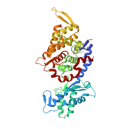
    7NKG

  • PubMed Abstract: 

    Methanogenic archaea operate an ancient, if not primordial, metabolic pathway that releases methane as an end-product. This last step is orchestrated by the methyl-coenzyme M reductase (MCR), which uses a nickel-containing F 430 -cofactor as the catalyst. MCR astounds the scientific world by its unique reaction chemistry, its numerous post-translational modifications, and its importance in biotechnology not only for production but also for capturing the greenhouse gas methane. In this report, we investigated MCR natively isolated from Methermicoccus shengliensis . This methanogen was isolated from a high-temperature oil reservoir and has recently been shown to convert lignin and coal derivatives into methane through a process called methoxydotrophic methanogenesis. A methoxydotrophic culture was obtained by growing M. shengliensis with 3,4,5-trimethoxybenzoate as the main carbon and energy source. Under these conditions, MCR represents more than 12% of the total protein content. The native MCR structure refined at a resolution of 1.6-Å precisely depicts the organization of a dimer of heterotrimers. Despite subtle surface remodeling and complete conservation of its active site with other homologues, MCR from the thermophile M. shengliensis contains the most limited number of post-translational modifications reported so far, questioning their physiological relevance in other relatives.


  • Organizational Affiliation

    Department of Microbiology, Institute for Water and Wetland Research, Radboud University, Heyendaalseweg 135, 6525 AJ Nijmegen, The Netherlands.


Macromolecules
Find similar proteins by:  (by identity cutoff)  |  3D Structure
Entity ID: 1
MoleculeChains Sequence LengthOrganismDetailsImage
Methyl-coenzyme M reductase alpha subunit
A, D, G, J
569Methermicoccus shengliensis DSM 18856Mutation(s): 0 
EC: 2.8.4.1
Entity Groups  
Sequence Clusters30% Identity50% Identity70% Identity90% Identity95% Identity100% Identity
Sequence Annotations
Expand
  • Reference Sequence
Find similar proteins by:  (by identity cutoff)  |  3D Structure
Entity ID: 2
MoleculeChains Sequence LengthOrganismDetailsImage
Methyl-coenzyme M reductase beta subunit
B, E, H, K
433Methermicoccus shengliensis DSM 18856Mutation(s): 0 
EC: 2.8.4.1
Entity Groups  
Sequence Clusters30% Identity50% Identity70% Identity90% Identity95% Identity100% Identity
Sequence Annotations
Expand
  • Reference Sequence
Find similar proteins by:  (by identity cutoff)  |  3D Structure
Entity ID: 3
MoleculeChains Sequence LengthOrganismDetailsImage
Methyl-coenzyme M reductase gamma subunit
C, F, I, L
248Methermicoccus shengliensis DSM 18856Mutation(s): 0 
EC: 2.8.4.1
Entity Groups  
Sequence Clusters30% Identity50% Identity70% Identity90% Identity95% Identity100% Identity
Sequence Annotations
Expand
  • Reference Sequence
Small Molecules
Ligands 6 Unique
IDChains Name / Formula / InChI Key2D Diagram3D Interactions
F43
Query on F43

Download Ideal Coordinates CCD File 
DA [auth G],
GA [auth J],
R [auth A],
V [auth D]
FACTOR 430
C42 H51 N6 Ni O13
XLFIRMYGVLUNOY-SXMZNAGASA-M
TP7
Query on TP7

Download Ideal Coordinates CCD File 
BA [auth G],
CA [auth G],
P [auth A],
Q [auth A]
Coenzyme B
C11 H22 N O7 P S
JBJSVEVEEGOEBZ-SCZZXKLOSA-N
COM
Query on COM

Download Ideal Coordinates CCD File 
AA [auth G],
FA [auth J],
O [auth A],
U [auth D]
1-THIOETHANESULFONIC ACID
C2 H6 O3 S2
ZNEWHQLOPFWXOF-UHFFFAOYSA-N
SO4
Query on SO4

Download Ideal Coordinates CCD File 
EA [auth H],
HA [auth K],
S [auth B],
W [auth E],
Y [auth E]
SULFATE ION
O4 S
QAOWNCQODCNURD-UHFFFAOYSA-L
GOL
Query on GOL

Download Ideal Coordinates CCD File 
M [auth A],
T [auth B],
X [auth E]
GLYCEROL
C3 H8 O3
PEDCQBHIVMGVHV-UHFFFAOYSA-N
K
Query on K

Download Ideal Coordinates CCD File 
N [auth A],
Z [auth G]
POTASSIUM ION
K
NPYPAHLBTDXSSS-UHFFFAOYSA-N
Modified Residues  3 Unique
IDChains TypeFormula2D DiagramParent
AGM
Query on AGM
A, D, G, J
L-PEPTIDE LINKINGC7 H17 N4 O2ARG
GL3
Query on GL3
A, D, G, J
L-PEPTIDE LINKINGC2 H5 N O SGLY
MHS
Query on MHS
A, D, G, J
L-PEPTIDE LINKINGC7 H11 N3 O2HIS
Experimental Data & Validation

Experimental Data

  • Method: X-RAY DIFFRACTION
  • Resolution: 1.60 Å
  • R-Value Free: 0.190 
  • R-Value Work: 0.172 
  • R-Value Observed: 0.173 
  • Space Group: P 21 21 21
Unit Cell:
Length ( Å )Angle ( ˚ )
a = 132.615α = 90
b = 148.177β = 90
c = 235.415γ = 90
Software Package:
Software NamePurpose
BUSTERrefinement
PDB_EXTRACTdata extraction
XDSdata reduction
SCALAdata scaling
PHASERphasing

Structure Validation

View Full Validation Report


Ligand Structure Quality Assessment 


Entry History & Funding Information

Deposition Data


Funding OrganizationLocationGrant Number
Max Planck SocietyGermany--
Netherlands Organisation for Scientific Research (NWO)NetherlandsGravitation grant 024.002.002
German Research Foundation (DFG)Germany3768/1-1

Revision History  (Full details and data files)

  • Version 1.0: 2021-05-19
    Type: Initial release
  • Version 1.1: 2024-01-31
    Changes: Data collection, Database references, Refinement description