7N63

X-ray structure of HCAN_0200, an aminotransferase from Helicobacter canadensis in complex with its external aldimine


Experimental Data Snapshot

  • Method: X-RAY DIFFRACTION
  • Resolution: 1.40 Å
  • R-Value Free: 0.190 
  • R-Value Work: 0.171 
  • R-Value Observed: 0.172 

wwPDB Validation   3D Report Full Report


Ligand Structure Quality Assessment 


This is version 1.3 of the entry. See complete history


Literature

Investigation of the enzymes required for the biosynthesis of an unusual formylated sugar in the emerging human pathogen Helicobacter canadensis.

Heisdorf, C.J.Griffiths, W.A.Thoden, J.B.Holden, H.M.

(2021) Protein Sci 30: 2144-2160

  • DOI: https://doi.org/10.1002/pro.4169
  • Primary Citation of Related Structures:  
    7N63, 7N67, 7N7A, 7N7B, 7N7C

  • PubMed Abstract: 

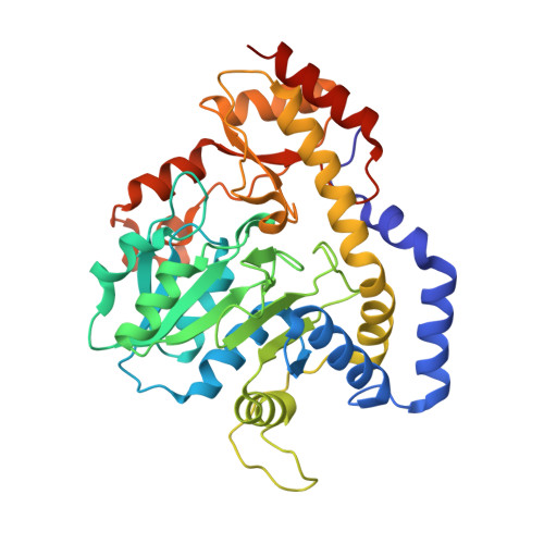
    It is now well established that the Gram-negative bacterium, Helicobacter pylori, causes gastritis in humans. In recent years, it has become apparent that the so-called non-pylori Helicobacters, normally infecting pigs, cats, and dogs, may also be involved in human pathology via zoonotic transmission. Indeed, more than 30 species of non-pylori Helicobacters have been identified thus far. One such organism is Helicobacter canadensis, an emerging pathogen whose genome sequence was published in 2009. Given our long-standing interest in the biosynthesis of N-formylated sugars found in the O-antigens of some Gram-negative bacteria, we were curious as to whether H. canadensis produces such unusual carbohydrates. Here, we demonstrate using both biochemical and structural techniques that the proteins encoded by the HCAN_0198, HCAN_0204, and HCAN_0200 genes in H. canadensis, correspond to a 3,4-ketoisomerase, a pyridoxal 5'-phosphate aminotransferase, and an N-formyltransferase, respectively. For this investigation, five high-resolution X-ray structures were determined and the kinetic parameters for the isomerase and the N-formyltransferase were measured. Based on these data, we suggest that the unusual sugar, 3-formamido-3,6-dideoxy-d-glucose, will most likely be found in the O-antigen of H. canadensis. Whether N-formylated sugars found in the O-antigen contribute to virulence is presently unclear, but it is intriguing that they have been observed in such pathogens as Francisella tularensis, Mycobacterium tuberculosis, and Brucella melitensis.


  • Organizational Affiliation

    Department of Biochemistry, University of Wisconsin, Madison, Wisconsin, USA.


Macromolecules
Find similar proteins by:  (by identity cutoff)  |  3D Structure
Entity ID: 1
MoleculeChains Sequence LengthOrganismDetailsImage
Putative aminotransferase369Helicobacter canadensis MIT 98-5491Mutation(s): 0 
Gene Names: HCAN_0204
UniProt
Find proteins for C5ZW06 (Helicobacter canadensis MIT 98-5491)
Explore C5ZW06 
Go to UniProtKB:  C5ZW06
Entity Groups  
Sequence Clusters30% Identity50% Identity70% Identity90% Identity95% Identity100% Identity
UniProt GroupC5ZW06
Sequence Annotations
Expand
  • Reference Sequence
Small Molecules
Ligands 4 Unique
IDChains Name / Formula / InChI Key2D Diagram3D Interactions
TQP (Subject of Investigation/LOI)
Query on TQP

Download Ideal Coordinates CCD File 
B [auth A](2R,3R,4S,5S,6R)-3,5-dihydroxy-4-{[(1E)-{3-hydroxy-2-methyl-5-[(phosphonooxy)methyl]pyridin-4-yl}methylidene]amino}-6-methyltetrahydro-2H-pyran-2-yl [(2R,3S,5R)-3-hydroxy-5-(5-methyl-2,4-dioxo-3,4-dihydropyrimidin-1(2H)-yl)tetrahydrofuran-2-yl]methyl dihydrogen diphosphate
C24 H35 N4 O19 P3
UJDWKFAFGYHSFM-HMSDFHDVSA-N
EDO
Query on EDO

Download Ideal Coordinates CCD File 
D [auth A]
E [auth A]
F [auth A]
G [auth A]
H [auth A]
D [auth A],
E [auth A],
F [auth A],
G [auth A],
H [auth A],
I [auth A]
1,2-ETHANEDIOL
C2 H6 O2
LYCAIKOWRPUZTN-UHFFFAOYSA-N
CL
Query on CL

Download Ideal Coordinates CCD File 
J [auth A]CHLORIDE ION
Cl
VEXZGXHMUGYJMC-UHFFFAOYSA-M
MG
Query on MG

Download Ideal Coordinates CCD File 
C [auth A]MAGNESIUM ION
Mg
JLVVSXFLKOJNIY-UHFFFAOYSA-N
Experimental Data & Validation

Experimental Data

  • Method: X-RAY DIFFRACTION
  • Resolution: 1.40 Å
  • R-Value Free: 0.190 
  • R-Value Work: 0.171 
  • R-Value Observed: 0.172 
  • Space Group: C 2 2 21
Unit Cell:
Length ( Å )Angle ( ˚ )
a = 61.005α = 90
b = 150.766β = 90
c = 91.058γ = 90
Software Package:
Software NamePurpose
REFMACrefinement
PDB_EXTRACTdata extraction
SAINTdata reduction
SADABSdata scaling
PHASERphasing

Structure Validation

View Full Validation Report


Ligand Structure Quality Assessment 


Entry History & Funding Information

Deposition Data


Funding OrganizationLocationGrant Number
National Institutes of Health/National Institute of General Medical Sciences (NIH/NIGMS)United StatesR35 GM134643

Revision History  (Full details and data files)

  • Version 1.0: 2021-06-23
    Type: Initial release
  • Version 1.1: 2021-08-25
    Changes: Database references
  • Version 1.2: 2021-09-22
    Changes: Database references
  • Version 1.3: 2023-10-18
    Changes: Data collection, Refinement description