7MFD

Autoinhibited BRAF:(14-3-3)2:MEK complex with the BRAF RBD resolved


Experimental Data Snapshot

  • Method: ELECTRON MICROSCOPY
  • Resolution: 3.66 Å
  • Aggregation State: PARTICLE 
  • Reconstruction Method: SINGLE PARTICLE 

wwPDB Validation   3D Report Full Report


This is version 1.1 of the entry. See complete history


Literature

Structural insights into the BRAF monomer-to-dimer transition mediated by RAS binding.

Martinez Fiesco, J.A.Durrant, D.E.Morrison, D.K.Zhang, P.

(2022) Nat Commun 13: 486-486

  • DOI: https://doi.org/10.1038/s41467-022-28084-3
  • Primary Citation of Related Structures:  
    7MFD, 7MFE, 7MFF

  • PubMed Abstract: 

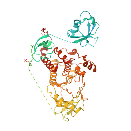
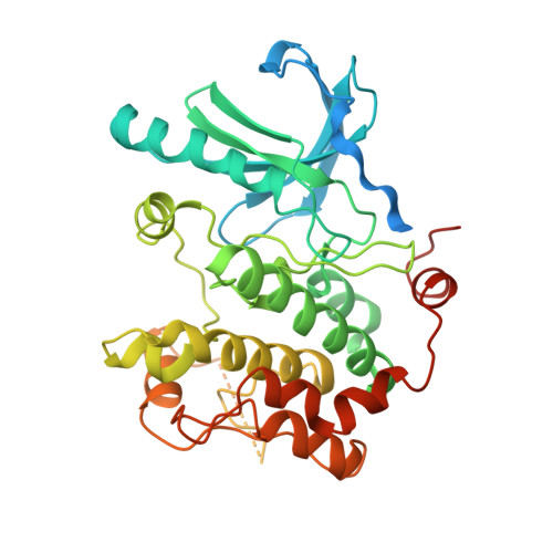
    RAF kinases are essential effectors of RAS, but how RAS binding initiates the conformational changes needed for autoinhibited RAF monomers to form active dimers has remained unclear. Here, we present cryo-electron microscopy structures of full-length BRAF complexes derived from mammalian cells: autoinhibited, monomeric BRAF:14-3-3 2 :MEK and BRAF:14-3-3 2 complexes, and an inhibitor-bound, dimeric BRAF 2 :14-3-3 2 complex, at 3.7, 4.1, and 3.9 Å resolution, respectively. In both autoinhibited, monomeric structures, the RAS binding domain (RBD) of BRAF is resolved, revealing that the RBD forms an extensive contact interface with the 14-3-3 protomer bound to the BRAF C-terminal site and that key basic residues required for RBD-RAS binding are exposed. Moreover, through structure-guided mutational studies, our findings indicate that RAS-RAF binding is a dynamic process and that RBD residues at the center of the RBD:14-3-3 interface have a dual function, first contributing to RAF autoinhibition and then to the full spectrum of RAS-RBD interactions.


  • Organizational Affiliation

    Center for Structural Biology, Center for Cancer Research, National Cancer Institute-Frederick, Frederick, MD, 21702, USA.


Macromolecules
Find similar proteins by:  (by identity cutoff)  |  3D Structure
Entity ID: 1
MoleculeChains Sequence LengthOrganismDetailsImage
Serine/threonine-protein kinase B-raf766Homo sapiensMutation(s): 0 
Gene Names: BRAFBRAF1RAFB1
EC: 2.7.11.1
UniProt & NIH Common Fund Data Resources
Find proteins for P15056 (Homo sapiens)
Explore P15056 
Go to UniProtKB:  P15056
PHAROS:  P15056
GTEx:  ENSG00000157764 
Entity Groups  
Sequence Clusters30% Identity50% Identity70% Identity90% Identity95% Identity100% Identity
UniProt GroupP15056
Sequence Annotations
Expand
  • Reference Sequence
Find similar proteins by:  (by identity cutoff)  |  3D Structure
Entity ID: 2
MoleculeChains Sequence LengthOrganismDetailsImage
Dual specificity mitogen-activated protein kinase kinase 1393Homo sapiensMutation(s): 0 
EC: 2.7.12.2
UniProt & NIH Common Fund Data Resources
Find proteins for Q02750 (Homo sapiens)
Explore Q02750 
Go to UniProtKB:  Q02750
PHAROS:  Q02750
GTEx:  ENSG00000169032 
Entity Groups  
Sequence Clusters30% Identity50% Identity70% Identity90% Identity95% Identity100% Identity
UniProt GroupQ02750
Sequence Annotations
Expand
  • Reference Sequence
Find similar proteins by:  (by identity cutoff)  |  3D Structure
Entity ID: 3
MoleculeChains Sequence LengthOrganismDetailsImage
14-3-3 protein zeta/delta
C, D
245Homo sapiensMutation(s): 0 
UniProt & NIH Common Fund Data Resources
Find proteins for P63104 (Homo sapiens)
Explore P63104 
Go to UniProtKB:  P63104
PHAROS:  P63104
GTEx:  ENSG00000164924 
Entity Groups  
Sequence Clusters30% Identity50% Identity70% Identity90% Identity95% Identity100% Identity
UniProt GroupP63104
Sequence Annotations
Expand
  • Reference Sequence
Small Molecules
Ligands 2 Unique
IDChains Name / Formula / InChI Key2D Diagram3D Interactions
CHU
Query on CHU

Download Ideal Coordinates CCD File 
G [auth B]N-(3-fluoro-4-{[4-methyl-2-oxo-7-(pyrimidin-2-yloxy)-2H-chromen-3-yl]methyl}pyridin-2-yl)-N'-methylsulfuric diamide
C21 H18 F N5 O5 S
LMMJFBMMJUMSJS-UHFFFAOYSA-N
ZN
Query on ZN

Download Ideal Coordinates CCD File 
E [auth A],
F [auth A]
ZINC ION
Zn
PTFCDOFLOPIGGS-UHFFFAOYSA-N
Modified Residues  1 Unique
IDChains TypeFormula2D DiagramParent
SEP
Query on SEP
A
L-PEPTIDE LINKINGC3 H8 N O6 PSER
Experimental Data & Validation

Experimental Data

  • Method: ELECTRON MICROSCOPY
  • Resolution: 3.66 Å
  • Aggregation State: PARTICLE 
  • Reconstruction Method: SINGLE PARTICLE 

Structure Validation

View Full Validation Report


Entry History & Funding Information

Deposition Data


Funding OrganizationLocationGrant Number
National Institutes of Health/National Cancer Institute (NIH/NCI)United StatesZIA BC 011744
National Institutes of Health/National Cancer Institute (NIH/NCI)United StatesZIA BC 010329

Revision History  (Full details and data files)

  • Version 1.0: 2022-01-26
    Type: Initial release
  • Version 1.1: 2022-02-09
    Changes: Database references