7L0J

Structure of AMH bound to AMHR2-ECD


Experimental Data Snapshot

  • Method: X-RAY DIFFRACTION
  • Resolution: 2.60 Å
  • R-Value Free: 0.268 
  • R-Value Work: 0.241 
  • R-Value Observed: 0.243 

wwPDB Validation   3D Report Full Report


Ligand Structure Quality Assessment 


This is version 1.2 of the entry. See complete history


Literature

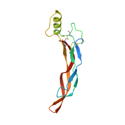
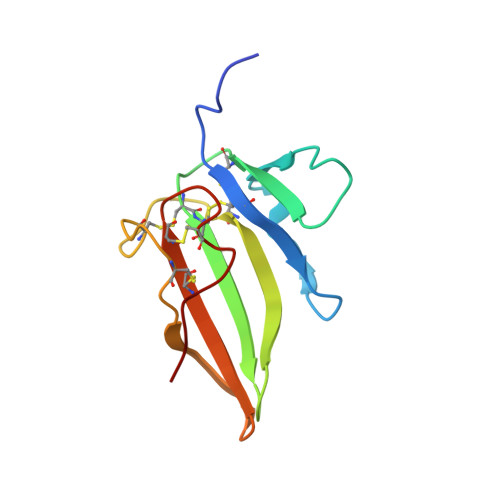
Structure of AMH bound to AMHR2 provides insight into a unique signaling pair in the TGF-beta family.

Hart, K.N.Stocker, W.A.Nagykery, N.G.Walton, K.L.Harrison, C.A.Donahoe, P.K.Pepin, D.Thompson, T.B.

(2021) Proc Natl Acad Sci U S A 118

  • DOI: https://doi.org/10.1073/pnas.2104809118
  • Primary Citation of Related Structures:  
    7L0J

  • PubMed Abstract: 

    Anti-Müllerian hormone (AMH), or Müllerian-inhibiting substance, is a protein hormone that promotes Müllerian duct regression during male fetal sexual differentiation and regulation of folliculogenesis in women. AMH is a member of the transforming growth factor beta (TGF-β) family, which has evolved to signal through its own dedicated type II receptor, AMH receptor type II (AMHR2). Structures of other TGF-β family members have revealed how ligands infer specificity for their cognate receptors; however, it is unknown how AMH binds AMHR2 at the molecular level. Therefore, in this study, we solved the X-ray crystal structure of AMH bound to the extracellular domain of AMHR2 to a resolution of 2.6Å. The structure reveals that while AMH binds AMHR2 in a similar location to Activin and BMP ligand binding to their type II receptors, differences in both AMH and AMHR2 account for a highly specific interaction. Furthermore, using an AMH responsive cell-based luciferase assay, we show that a conformation in finger 1 of AMHR2 and a salt bridge formed by K534 on AMH and D81/E84 of AMHR2 are key to the AMH/AMHR2 interaction. Overall, our study highlights how AMH engages AMHR2 using a modified paradigm of receptor binding facilitated by modifications to the three-finger toxin fold of AMHR2. Furthermore, understanding these elements contributing to the specificity of binding will help in the design of agonists or antagonists or the selection of antibody therapies.


  • Organizational Affiliation

    Department of Pharmacology and Systems Physiology, University of Cincinnati, Cincinnati, OH 45267.


Macromolecules
Find similar proteins by:  (by identity cutoff)  |  3D Structure
Entity ID: 1
MoleculeChains Sequence LengthOrganismDetailsImage
Muellerian-inhibiting factor109Homo sapiensMutation(s): 1 
Gene Names: AMHMIF
UniProt & NIH Common Fund Data Resources
Find proteins for P03971 (Homo sapiens)
Explore P03971 
Go to UniProtKB:  P03971
PHAROS:  P03971
GTEx:  ENSG00000104899 
Entity Groups  
Sequence Clusters30% Identity50% Identity70% Identity90% Identity95% Identity100% Identity
UniProt GroupP03971
Sequence Annotations
Expand
  • Reference Sequence
Find similar proteins by:  (by identity cutoff)  |  3D Structure
Entity ID: 2
MoleculeChains Sequence LengthOrganismDetailsImage
Anti-Muellerian hormone type-2 receptor111Homo sapiensMutation(s): 0 
Gene Names: AMHR2AMHRMISR2
EC: 2.7.11.30
UniProt & NIH Common Fund Data Resources
Find proteins for Q16671 (Homo sapiens)
Explore Q16671 
Go to UniProtKB:  Q16671
PHAROS:  Q16671
GTEx:  ENSG00000135409 
Entity Groups  
Sequence Clusters30% Identity50% Identity70% Identity90% Identity95% Identity100% Identity
UniProt GroupQ16671
Sequence Annotations
Expand
  • Reference Sequence
Small Molecules
Ligands 2 Unique
IDChains Name / Formula / InChI Key2D Diagram3D Interactions
NAG
Query on NAG

Download Ideal Coordinates CCD File 
C [auth B]2-acetamido-2-deoxy-beta-D-glucopyranose
C8 H15 N O6
OVRNDRQMDRJTHS-FMDGEEDCSA-N
SO4
Query on SO4

Download Ideal Coordinates CCD File 
D [auth B]SULFATE ION
O4 S
QAOWNCQODCNURD-UHFFFAOYSA-L
Experimental Data & Validation

Experimental Data

  • Method: X-RAY DIFFRACTION
  • Resolution: 2.60 Å
  • R-Value Free: 0.268 
  • R-Value Work: 0.241 
  • R-Value Observed: 0.243 
  • Space Group: P 41 21 2
Unit Cell:
Length ( Å )Angle ( ˚ )
a = 66.376α = 90
b = 66.376β = 90
c = 125.922γ = 90
Software Package:
Software NamePurpose
PHENIXrefinement
DIALSdata reduction
Aimlessdata scaling
PHASERphasing

Structure Validation

View Full Validation Report


Ligand Structure Quality Assessment 


Entry History & Funding Information

Deposition Data


Funding OrganizationLocationGrant Number
National Institutes of Health/National Institute of Environmental Health Sciences (NIH/NIEHS)United States5T32ES007250-30

Revision History  (Full details and data files)

  • Version 1.0: 2021-06-23
    Type: Initial release
  • Version 1.1: 2021-07-28
    Changes: Database references
  • Version 1.2: 2023-10-18
    Changes: Data collection, Database references, Refinement description