7K88

Crystal structure of bovine RPE65 in complex with hexaethylene glycol monooctyl ether


Experimental Data Snapshot

  • Method: X-RAY DIFFRACTION
  • Resolution: 2.10 Å
  • R-Value Free: 0.220 
  • R-Value Work: 0.189 
  • R-Value Observed: 0.191 

wwPDB Validation   3D Report Full Report


Ligand Structure Quality Assessment 


This is version 1.2 of the entry. See complete history


Literature

Rational Alteration of Pharmacokinetics of Chiral Fluorinated and Deuterated Derivatives of Emixustat for Retinal Therapy.

Blum, E.Zhang, J.Zaluski, J.Einstein, D.E.Korshin, E.E.Kubas, A.Gruzman, A.Tochtrop, G.P.Kiser, P.D.Palczewski, K.

(2021) J Med Chem 64: 8287-8302

  • DOI: https://doi.org/10.1021/acs.jmedchem.1c00279
  • Primary Citation of Related Structures:  
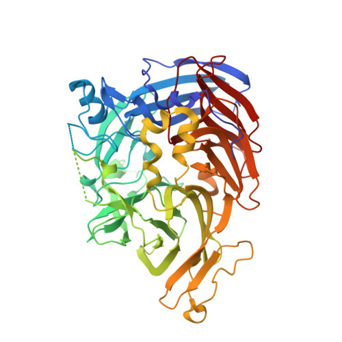
    7K88, 7K89, 7K8G, 7L0E

  • PubMed Abstract: 

    Recycling of all- trans -retinal to 11- cis -retinal through the visual cycle is a fundamental metabolic pathway in the eye. A potent retinoid isomerase (RPE65) inhibitor, ( R )-emixustat, has been developed and tested in several clinical trials; however, it has not received regulatory approval for use in any specific retinopathy. Rapid clearance of this drug presents challenges to maintaining concentrations in eyes within a therapeutic window. To address this pharmacokinetic inadequacy, we rationally designed and synthesized a series of emixustat derivatives with strategically placed fluorine and deuterium atoms to slow down the key metabolic transformations known for emixustat. Crystal structures and quantum chemical analysis of RPE65 in complex with the most potent emixustat derivatives revealed the structural and electronic bases for how fluoro substituents can be favorably accommodated within the active site pocket of RPE65. We found a close (∼3.0 Å) F-π interaction that is predicted to contribute ∼2.4 kcal/mol to the overall binding energy.


  • Organizational Affiliation

    Department of Chemistry, Faculty of Exact Sciences, Bar-Ilan University, Ramat-Gan 5290002, Israel.


Macromolecules
Find similar proteins by:  (by identity cutoff)  |  3D Structure
Entity ID: 1
MoleculeChains Sequence LengthOrganismDetailsImage
Retinoid isomerohydrolase
A, B
533Bos taurusMutation(s): 1 
EC: 3.1.1.64 (PDB Primary Data), 5.3.3.22 (PDB Primary Data)
UniProt
Find proteins for Q28175 (Bos taurus)
Explore Q28175 
Go to UniProtKB:  Q28175
Entity Groups  
Sequence Clusters30% Identity50% Identity70% Identity90% Identity95% Identity100% Identity
UniProt GroupQ28175
Sequence Annotations
Expand
  • Reference Sequence
Small Molecules
Ligands 4 Unique
IDChains Name / Formula / InChI Key2D Diagram3D Interactions
32M (Subject of Investigation/LOI)
Query on 32M

Download Ideal Coordinates CCD File 
D [auth A],
F [auth B]
3,6,9,12,15,18-hexaoxahexacosan-1-ol
C20 H42 O7
VVSOZSGWKFPDFX-UHFFFAOYSA-N
PG4
Query on PG4

Download Ideal Coordinates CCD File 
G [auth B]TETRAETHYLENE GLYCOL
C8 H18 O5
UWHCKJMYHZGTIT-UHFFFAOYSA-N
FE2 (Subject of Investigation/LOI)
Query on FE2

Download Ideal Coordinates CCD File 
C [auth A],
E [auth B]
FE (II) ION
Fe
CWYNVVGOOAEACU-UHFFFAOYSA-N
NA
Query on NA

Download Ideal Coordinates CCD File 
H [auth B]SODIUM ION
Na
FKNQFGJONOIPTF-UHFFFAOYSA-N
Experimental Data & Validation

Experimental Data

  • Method: X-RAY DIFFRACTION
  • Resolution: 2.10 Å
  • R-Value Free: 0.220 
  • R-Value Work: 0.189 
  • R-Value Observed: 0.191 
  • Space Group: P 65
Unit Cell:
Length ( Å )Angle ( ˚ )
a = 175.917α = 90
b = 175.917β = 90
c = 86.314γ = 120
Software Package:
Software NamePurpose
REFMACrefinement
XDSdata reduction
XDSdata scaling
REFMACphasing

Structure Validation

View Full Validation Report


Ligand Structure Quality Assessment 


Entry History 

Deposition Data

  • Released Date: 2021-06-02 
  • Deposition Author(s): Kiser, P.D.

Revision History  (Full details and data files)

  • Version 1.0: 2021-06-02
    Type: Initial release
  • Version 1.1: 2021-07-07
    Changes: Database references
  • Version 1.2: 2023-10-18
    Changes: Data collection, Database references, Refinement description