7JM7

Structure of human CLC-7/OSTM1 complex

  • Classification: MEMBRANE PROTEIN
  • Organism(s): Homo sapiens
  • Expression System: Homo sapiens
  • Mutation(s): No 
  • Membrane Protein: Yes  OPMPDBTMMemProtMD

  • Deposited: 2020-07-31 Released: 2020-09-02 
  • Deposition Author(s): Schrecker, M., Hite, R.
  • Funding Organization(s): National Institutes of Health/National Institute of General Medical Sciences (NIH/NIGMS), National Institutes of Health/National Cancer Institute (NIH/NCI)

Experimental Data Snapshot

  • Method: ELECTRON MICROSCOPY
  • Resolution: 2.82 Å
  • Aggregation State: PARTICLE 
  • Reconstruction Method: SINGLE PARTICLE 

wwPDB Validation   3D Report Full Report


This is version 1.0 of the entry. See complete history


Literature

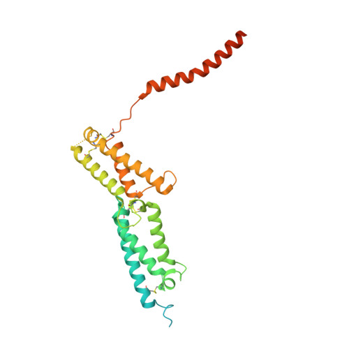
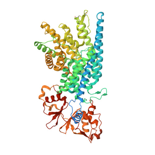
Cryo-EM structure of the lysosomal chloride-proton exchanger CLC-7 in complex with OSTM1.

Schrecker, M.Korobenko, J.Hite, R.K.

(2020) Elife 9

  • DOI: https://doi.org/10.7554/eLife.59555
  • Primary Citation of Related Structures:  
    7JM6, 7JM7

  • PubMed Abstract: 

    The chloride-proton exchanger CLC-7 plays critical roles in lysosomal homeostasis and bone regeneration and its mutation can lead to osteopetrosis, lysosomal storage disease and neurological disorders. In lysosomes and the ruffled border of osteoclasts, CLC-7 requires a β-subunit, OSTM1, for stability and activity. Here, we present electron cryomicroscopy structures of CLC-7 in occluded states by itself and in complex with OSTM1, determined at resolutions up to 2.8 Å. In the complex, the luminal surface of CLC-7 is entirely covered by a dimer of the heavily glycosylated and disulfide-bonded OSTM1, which serves to protect CLC-7 from the degradative environment of the lysosomal lumen. OSTM1 binding does not induce large-scale rearrangements of CLC-7, but does have minor effects on the conformation of the ion-conduction pathway, potentially contributing to its regulatory role. These studies provide insights into the role of OSTM1 and serve as a foundation for understanding the mechanisms of CLC-7 regulation.


  • Organizational Affiliation

    Structural Biology Program, Memorial Sloan Kettering Cancer Center, New York, United States.


Macromolecules
Find similar proteins by:  (by identity cutoff)  |  3D Structure
Entity ID: 1
MoleculeChains Sequence LengthOrganismDetailsImage
Osteopetrosis-associated transmembrane protein 1A [auth B],
C [auth D]
334Homo sapiensMutation(s): 0 
Gene Names: OSTM1GLHSPC019UNQ6098/PRO21201
Membrane Entity: Yes 
UniProt & NIH Common Fund Data Resources
Find proteins for Q86WC4 (Homo sapiens)
Explore Q86WC4 
Go to UniProtKB:  Q86WC4
PHAROS:  Q86WC4
GTEx:  ENSG00000081087 
Entity Groups  
Sequence Clusters30% Identity50% Identity70% Identity90% Identity95% Identity100% Identity
UniProt GroupQ86WC4
Sequence Annotations
Expand
  • Reference Sequence
Find similar proteins by:  (by identity cutoff)  |  3D Structure
Entity ID: 2
MoleculeChains Sequence LengthOrganismDetailsImage
H(+)/Cl(-) exchange transporter 7B [auth A],
D [auth C]
805Homo sapiensMutation(s): 0 
Gene Names: CLCN7
Membrane Entity: Yes 
UniProt & NIH Common Fund Data Resources
Find proteins for P51798 (Homo sapiens)
Explore P51798 
Go to UniProtKB:  P51798
PHAROS:  P51798
GTEx:  ENSG00000103249 
Entity Groups  
Sequence Clusters30% Identity50% Identity70% Identity90% Identity95% Identity100% Identity
UniProt GroupP51798
Sequence Annotations
Expand
  • Reference Sequence
Oligosaccharides

Help

Entity ID: 3
MoleculeChains Length2D Diagram Glycosylation3D Interactions
alpha-D-mannopyranose-(1-3)-[alpha-D-mannopyranose-(1-6)]beta-D-mannopyranose-(1-4)-2-acetamido-2-deoxy-beta-D-glucopyranose-(1-4)-2-acetamido-2-deoxy-beta-D-glucopyranose
E, H
5N/A
Glycosylation Resources
GlyTouCan:  G22768VO
GlyCosmos:  G22768VO
GlyGen:  G22768VO
Entity ID: 4
MoleculeChains Length2D Diagram Glycosylation3D Interactions
2-acetamido-2-deoxy-beta-D-glucopyranose-(1-4)-2-acetamido-2-deoxy-beta-D-glucopyranose
F, G, I, J
2N-Glycosylation
Glycosylation Resources
GlyTouCan:  G42666HT
GlyCosmos:  G42666HT
GlyGen:  G42666HT
Small Molecules
Ligands 5 Unique
IDChains Name / Formula / InChI Key2D Diagram3D Interactions
0J1 (Subject of Investigation/LOI)
Query on 0J1

Download Ideal Coordinates CCD File 
DA [auth C],
T [auth A]
(2R)-3-{[(R)-hydroxy{[(1S,2R,3S,4S,5R,6R)-2,3,4,6-tetrahydroxy-5-(phosphonooxy)cyclohexyl]oxy}phosphoryl]oxy}propane-1,2-diyl dinonanoate
C27 H52 O16 P2
IELCLCXLJSIZNX-ZAIPYKEWSA-N
ATP
Query on ATP

Download Ideal Coordinates CCD File 
O [auth A],
Y [auth C]
ADENOSINE-5'-TRIPHOSPHATE
C10 H16 N5 O13 P3
ZKHQWZAMYRWXGA-KQYNXXCUSA-N
NAG (Subject of Investigation/LOI)
Query on NAG

Download Ideal Coordinates CCD File 
K [auth B]
L [auth B]
M [auth B]
N [auth B]
U [auth D]
K [auth B],
L [auth B],
M [auth B],
N [auth B],
U [auth D],
V [auth D],
W [auth D],
X [auth D]
2-acetamido-2-deoxy-beta-D-glucopyranose
C8 H15 N O6
OVRNDRQMDRJTHS-FMDGEEDCSA-N
CL
Query on CL

Download Ideal Coordinates CCD File 
AA [auth C]
BA [auth C]
CA [auth C]
Q [auth A]
R [auth A]
AA [auth C],
BA [auth C],
CA [auth C],
Q [auth A],
R [auth A],
S [auth A]
CHLORIDE ION
Cl
VEXZGXHMUGYJMC-UHFFFAOYSA-M
MG
Query on MG

Download Ideal Coordinates CCD File 
P [auth A],
Z [auth C]
MAGNESIUM ION
Mg
JLVVSXFLKOJNIY-UHFFFAOYSA-N
Experimental Data & Validation

Experimental Data

  • Method: ELECTRON MICROSCOPY
  • Resolution: 2.82 Å
  • Aggregation State: PARTICLE 
  • Reconstruction Method: SINGLE PARTICLE 
EM Software:
TaskSoftware PackageVersion
MODEL REFINEMENTPHENIX
RECONSTRUCTIONcryoSPARC

Structure Validation

View Full Validation Report


Entry History & Funding Information

Deposition Data


Funding OrganizationLocationGrant Number
National Institutes of Health/National Institute of General Medical Sciences (NIH/NIGMS)United StatesGM103310
National Institutes of Health/National Cancer Institute (NIH/NCI)United StatesP30 CA008748

Revision History  (Full details and data files)

  • Version 1.0: 2020-09-02
    Type: Initial release