7F83

Crystal Structure of a receptor in Complex with inverse agonist


Experimental Data Snapshot

  • Method: X-RAY DIFFRACTION
  • Resolution: 2.94 Å
  • R-Value Free: 0.265 
  • R-Value Work: 0.227 
  • R-Value Observed: 0.229 

wwPDB Validation   3D Report Full Report


Ligand Structure Quality Assessment 


This is version 1.2 of the entry. See complete history


Literature

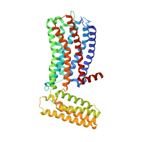
Molecular mechanism of agonism and inverse agonism in ghrelin receptor.

Qin, J.Cai, Y.Xu, Z.Ming, Q.Ji, S.Y.Wu, C.Zhang, H.Mao, C.Shen, D.D.Hirata, K.Ma, Y.Yan, W.Zhang, Y.Shao, Z.

(2022) Nat Commun 13: 300-300

  • DOI: https://doi.org/10.1038/s41467-022-27975-9
  • Primary Citation of Related Structures:  
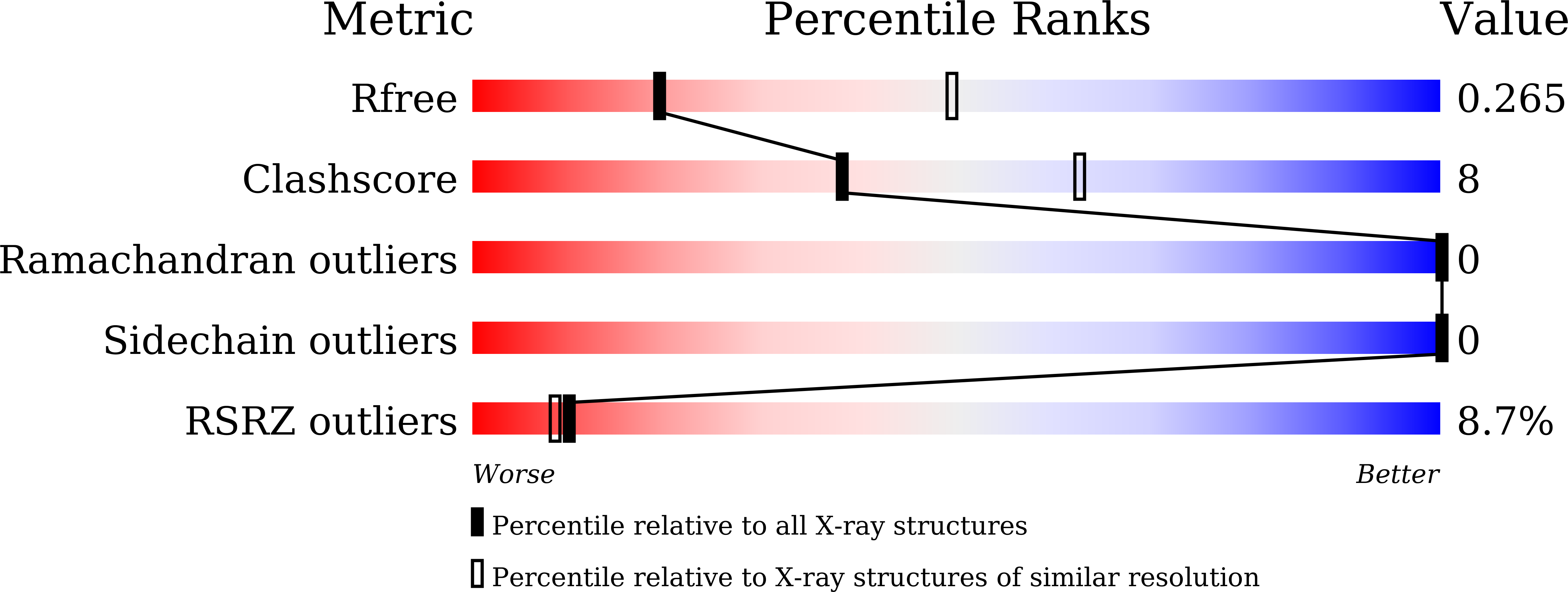
    7F83, 7W2Z

  • PubMed Abstract: 

    Much effort has been invested in the investigation of the structural basis of G protein-coupled receptors (GPCRs) activation. Inverse agonists, which can inhibit GPCRs with constitutive activity, are considered useful therapeutic agents, but the molecular mechanism of such ligands remains insufficiently understood. Here, we report a crystal structure of the ghrelin receptor bound to the inverse agonist PF-05190457 and a cryo-electron microscopy structure of the active ghrelin receptor-Go complex bound to the endogenous agonist ghrelin. Our structures reveal a distinct binding mode of the inverse agonist PF-05190457 in the ghrelin receptor, different from the binding mode of agonists and neutral antagonists. Combining the structural comparisons and cellular function assays, we find that a polar network and a notable hydrophobic cluster are required for receptor activation and constitutive activity. Together, our study provides insights into the detailed mechanism of ghrelin receptor binding to agonists and inverse agonists, and paves the way to design specific ligands targeting ghrelin receptors.


  • Organizational Affiliation

    Department of Biophysics and Department of Pathology of Sir Run Run Shaw Hospital, Zhejiang University School of Medicine, 310058, Hangzhou, China.


Macromolecules
Find similar proteins by:  (by identity cutoff)  |  3D Structure
Entity ID: 1
MoleculeChains Sequence LengthOrganismDetailsImage
Growth hormone secretagogue receptor type 1,Soluble cytochrome b562
A, B
416Homo sapiensEscherichia coliMutation(s): 5 
Gene Names: GHSRcybC
Membrane Entity: Yes 
UniProt & NIH Common Fund Data Resources
Find proteins for Q92847 (Homo sapiens)
Explore Q92847 
Go to UniProtKB:  Q92847
PHAROS:  Q92847
GTEx:  ENSG00000121853 
Find proteins for P0ABE7 (Escherichia coli)
Explore P0ABE7 
Go to UniProtKB:  P0ABE7
Entity Groups  
Sequence Clusters30% Identity50% Identity70% Identity90% Identity95% Identity100% Identity
UniProt GroupsP0ABE7Q92847
Sequence Annotations
Expand
  • Reference Sequence
Small Molecules
Ligands 2 Unique
IDChains Name / Formula / InChI Key2D Diagram3D Interactions
1KQ
Query on 1KQ

Download Ideal Coordinates CCD File 
E [auth A],
J [auth B]
2-(2-methylimidazo[2,1-b][1,3]thiazol-6-yl)-1-[2-[(1R)-5-(6-methylpyrimidin-4-yl)-2,3-dihydro-1H-inden-1-yl]-2,7-diazaspiro[3.5]nonan-7-yl]ethanone
C29 H32 N6 O S
ZIUDADZJCKGWKR-AREMUKBSSA-N
OLC
Query on OLC

Download Ideal Coordinates CCD File 
C [auth A]
D [auth A]
F [auth B]
G [auth B]
H [auth B]
C [auth A],
D [auth A],
F [auth B],
G [auth B],
H [auth B],
I [auth B]
(2R)-2,3-dihydroxypropyl (9Z)-octadec-9-enoate
C21 H40 O4
RZRNAYUHWVFMIP-GDCKJWNLSA-N
Experimental Data & Validation

Experimental Data

  • Method: X-RAY DIFFRACTION
  • Resolution: 2.94 Å
  • R-Value Free: 0.265 
  • R-Value Work: 0.227 
  • R-Value Observed: 0.229 
  • Space Group: P 1 21 1
Unit Cell:
Length ( Å )Angle ( ˚ )
a = 84.71α = 90
b = 58.68β = 90.6
c = 119.24γ = 90
Software Package:
Software NamePurpose
PHENIXrefinement
XDSdata reduction
XSCALEdata scaling
PDB_EXTRACTdata extraction
PHENIXphasing

Structure Validation

View Full Validation Report


Ligand Structure Quality Assessment 


Entry History 

Deposition Data

Revision History  (Full details and data files)

  • Version 1.0: 2022-01-19
    Type: Initial release
  • Version 1.1: 2022-01-26
    Changes: Database references
  • Version 1.2: 2023-11-29
    Changes: Data collection, Refinement description