


Growth-Coupled Evolutionary Pressure Improving Epimerases for D-Allulose Biosynthesis Using a Biosensor-Assisted In Vivo Selection Platform
Li, C., Gao, H., Li, H., Wang, T., Lu, F.P., Qin, H.-M.(2024) Adv Sci (Weinh)
Experimental Data Snapshot
Entity ID: 1 | |||||
|---|---|---|---|---|---|
| Molecule | Chains | Sequence Length | Organism | Details | Image |
| D-tagatose 3-epimerase | 284 | Agrobacterium sp. SUL3 | Mutation(s): 0 Gene Names: AKG12_23230 EC: 5.1.3 | 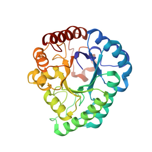 | |
Entity Groups | |||||
| Sequence Clusters | 30% Identity50% Identity70% Identity90% Identity95% Identity100% Identity | ||||
Sequence AnnotationsExpand | |||||
| |||||
| Ligands 2 Unique | |||||
|---|---|---|---|---|---|
| ID | Chains | Name / Formula / InChI Key | 2D Diagram | 3D Interactions | |
| PSJ (Subject of Investigation/LOI) Query on PSJ | E [auth A], G [auth B], I [auth C], K [auth D] | D-psicose C6 H12 O6 BJHIKXHVCXFQLS-PUFIMZNGSA-N |  | ||
| MG (Subject of Investigation/LOI) Query on MG | F [auth A], H [auth B], J [auth C], L [auth D] | MAGNESIUM ION Mg JLVVSXFLKOJNIY-UHFFFAOYSA-N |  | ||
| Length ( Å ) | Angle ( ˚ ) |
|---|---|
| a = 66.173 | α = 90 |
| b = 97.517 | β = 90 |
| c = 159.922 | γ = 90 |
| Software Name | Purpose |
|---|---|
| PHENIX | refinement |
| XDS | data reduction |
| Aimless | data scaling |
| PHASER | phasing |
RCSB PDB (citation) is hosted by
RCSB PDB is a member of the

