7D6Q

Crystal structure of the Stx2a


Experimental Data Snapshot

  • Method: X-RAY DIFFRACTION
  • Resolution: 1.80 Å
  • R-Value Free: 0.190 
  • R-Value Work: 0.163 
  • R-Value Observed: 0.164 

wwPDB Validation   3D Report Full Report


Ligand Structure Quality Assessment 


This is version 1.2 of the entry. See complete history


Literature

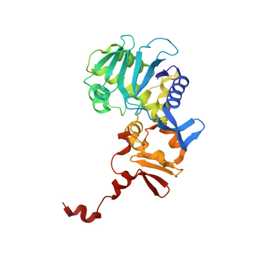
Identification of a peptide motif that potently inhibits two functionally distinct subunits of Shiga toxin.

Watanabe-Takahashi, M.Tamada, M.Senda, M.Hibino, M.Shimizu, E.Okuta, A.Miyazawa, A.Senda, T.Nishikawa, K.

(2021) Commun Biol 4: 538-538

  • DOI: https://doi.org/10.1038/s42003-021-02068-3
  • Primary Citation of Related Structures:  
    7D6Q, 7D6R

  • PubMed Abstract: 

    Shiga toxin (Stx) is a major virulence factor of enterohemorrhagic Escherichia coli, which causes fatal systemic complications. Here, we identified a tetravalent peptide that inhibited Stx by targeting its receptor-binding, B-subunit pentamer through a multivalent interaction. A monomeric peptide with the same motif, however, did not bind to the B-subunit pentamer. Instead, the monomer inhibited cytotoxicity with remarkable potency by binding to the catalytic A-subunit. An X-ray crystal structure analysis to 1.6 Å resolution revealed that the monomeric peptide fully occupied the catalytic cavity, interacting with Glu167 and Arg170, both of which are essential for catalytic activity. Thus, the peptide motif demonstrated potent inhibition of two functionally distinct subunits of Stx.


  • Organizational Affiliation

    Department of Molecular Life Sciences, Graduate School of Life and Medical Sciences, Doshisha University, Kyoto, Japan.


Macromolecules
Find similar proteins by:  (by identity cutoff)  |  3D Structure
Entity ID: 1
MoleculeChains Sequence LengthOrganismDetailsImage
rRNA N-glycosylase297Escherichia coliMutation(s): 0 
Gene Names: stx2a
EC: 3.2.2.22
UniProt
Find proteins for Q8XBV2 (Escherichia coli)
Explore Q8XBV2 
Go to UniProtKB:  Q8XBV2
Entity Groups  
Sequence Clusters30% Identity50% Identity70% Identity90% Identity95% Identity100% Identity
UniProt GroupQ8XBV2
Sequence Annotations
Expand
  • Reference Sequence
Find similar proteins by:  (by identity cutoff)  |  3D Structure
Entity ID: 2
MoleculeChains Sequence LengthOrganismDetailsImage
Shiga toxin 2 B subunit
B, C, D, E, F
70Escherichia coliMutation(s): 0 
Gene Names: stxIIstx2Bstx2B_2stx2dBstx2vBstxB2vtx2B
UniProt
Find proteins for Q7DJJ2 (Escherichia coli)
Explore Q7DJJ2 
Go to UniProtKB:  Q7DJJ2
Entity Groups  
Sequence Clusters30% Identity50% Identity70% Identity90% Identity95% Identity100% Identity
UniProt GroupQ7DJJ2
Sequence Annotations
Expand
  • Reference Sequence
Experimental Data & Validation

Experimental Data

  • Method: X-RAY DIFFRACTION
  • Resolution: 1.80 Å
  • R-Value Free: 0.190 
  • R-Value Work: 0.163 
  • R-Value Observed: 0.164 
  • Space Group: P 61
Unit Cell:
Length ( Å )Angle ( ˚ )
a = 146.695α = 90
b = 146.695β = 90
c = 60.875γ = 120
Software Package:
Software NamePurpose
XDSdata reduction
XSCALEdata scaling
Cootmodel building
MOLREPphasing
PHENIXrefinement

Structure Validation

View Full Validation Report


Ligand Structure Quality Assessment 


Entry History & Funding Information

Deposition Data


Funding OrganizationLocationGrant Number
Japan Agency for Medical Research and Development (AMED)JapanJP19am0101001

Revision History  (Full details and data files)

  • Version 1.0: 2021-04-14
    Type: Initial release
  • Version 1.1: 2021-05-26
    Changes: Database references
  • Version 1.2: 2023-11-29
    Changes: Data collection, Database references, Refinement description