7D6F

The crystal structure of ARMS-PBM/MAGI2-PDZ4


Experimental Data Snapshot

  • Method: X-RAY DIFFRACTION
  • Resolution: 3.00 Å
  • R-Value Free: 0.281 
  • R-Value Work: 0.235 
  • R-Value Observed: 0.238 

wwPDB Validation   3D Report Full Report


Ligand Structure Quality Assessment 


This is version 2.2 of the entry. See complete history


Literature

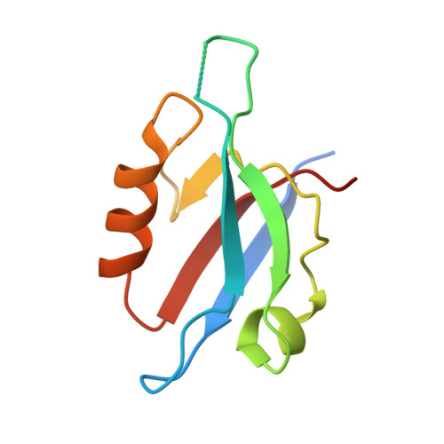
Crystal structure of the PDZ4 domain of MAGI2 in complex with PBM of ARMS reveals a canonical PDZ recognition mode.

Zhang, Y.Zhong, Z.Ye, J.Wang, C.

(2021) Neurochem Int 149: 105152-105152

  • DOI: https://doi.org/10.1016/j.neuint.2021.105152
  • Primary Citation of Related Structures:  
    7D6F

  • PubMed Abstract: 

    Membrane-associated guanylate kinase, WW and PDZ domain-containing protein 2 (MAGI2) is a neuronal scaffold protein that plays critical roles at synaptic junctions by assembling neurotransmitter receptors and cell adhesion proteins through its multiple protein-protein interaction domains, including six PDZ domains, two phosphoserine-phosphothreonine binding WW domains, and a guanylate kinase GK domain. Previous studies showed that MAGI2 participates in formation of tetrameric complexes with PDZ-GEF1, TrkA receptor, and ankyrin repeat-rich membrane spanning (ARMS) protein at late endosomes and is crucial for neurite outgrowth. However, the molecular mechanism governing the assembly of these complexes remains unknown. Here, we characterize the direct interaction between MAGI2 and ARMS through multiple biochemical assays. Moreover, our solved crystal structure of the truncated PDZ4/PBM (PDZ binding motifs) complex of MAGI2 and ARMS proteins (MAGI2-PDZ4/ARMS-PBM) reveals that the binding interface lies between the αB/βB groove from the PDZ4 of MAGI2 and the C-terminal PBM from ARMS. The structure reveals high similarity to others in this protein family where canonical PDZ/PBM interactions are observed. However, the conserved "GLGF" motif in the PSD-95-PDZ3 changes to "GFGF" in the MAGI2-PDZ4/ARMS-PBM complex. We further validated our crystal structure through serial mutagenesis assays. Taken together, our study provides the biochemical details and binding mechanisms that underpin the stabilization of the MAGI2-PDZ4/ARMS-PBM complex, thereby offering a biochemical and structural basis for further understanding of the functional roles of MAGI2, ARMS, PDZ-GEF1, and TrkA in forming the tetrameric receptor complex in neuronal signaling.


  • Organizational Affiliation

    Department of Neurology, The First Affiliated Hospital of USTC, Division of Life Sciences and Medicine, MOE Key Laboratory for Cellular Dynamics, Hefei National Laboratory for Physical Sciences at the Microscale, University of Science and Technology of China, 230001, Hefei, China.


Macromolecules
Find similar proteins by:  (by identity cutoff)  |  3D Structure
Entity ID: 1
MoleculeChains Sequence LengthOrganismDetailsImage
Membrane-associated guanylate kinase, WW and PDZ domain-containing protein 2101Mus musculusMutation(s): 0 
Gene Names: Magi2Acvrinp1Aip1Arip1
UniProt & NIH Common Fund Data Resources
Find proteins for Q9WVQ1 (Mus musculus)
Explore Q9WVQ1 
Go to UniProtKB:  Q9WVQ1
IMPC:  MGI:1354953
Entity Groups  
Sequence Clusters30% Identity50% Identity70% Identity90% Identity95% Identity100% Identity
UniProt GroupQ9WVQ1
Sequence Annotations
Expand
  • Reference Sequence

Find similar proteins by:  Sequence   |   3D Structure  

Entity ID: 2
MoleculeChains Sequence LengthOrganismDetailsImage
Kinase D-interacting substrate of 220 kDa19Rattus norvegicusMutation(s): 0 
Gene Names: Kidins220Arms
UniProt
Find proteins for Q9EQG6 (Rattus norvegicus)
Explore Q9EQG6 
Go to UniProtKB:  Q9EQG6
Entity Groups  
Sequence Clusters30% Identity50% Identity70% Identity90% Identity95% Identity100% Identity
UniProt GroupQ9EQG6
Sequence Annotations
Expand
  • Reference Sequence
Small Molecules
Ligands 1 Unique
IDChains Name / Formula / InChI Key2D Diagram3D Interactions
GOL (Subject of Investigation/LOI)
Query on GOL

Download Ideal Coordinates CCD File 
C [auth A]GLYCEROL
C3 H8 O3
PEDCQBHIVMGVHV-UHFFFAOYSA-N
Experimental Data & Validation

Experimental Data

  • Method: X-RAY DIFFRACTION
  • Resolution: 3.00 Å
  • R-Value Free: 0.281 
  • R-Value Work: 0.235 
  • R-Value Observed: 0.238 
  • Space Group: C 2 2 2
Unit Cell:
Length ( Å )Angle ( ˚ )
a = 74.045α = 90
b = 95.666β = 90
c = 35.167γ = 90
Software Package:
Software NamePurpose
PHENIXrefinement
HKL-2000data reduction
HKL-2000data scaling
PDB_EXTRACTdata extraction
MOLREPphasing

Structure Validation

View Full Validation Report


Ligand Structure Quality Assessment 


Entry History 

Deposition Data

Revision History  (Full details and data files)

  • Version 1.0: 2020-11-04
    Type: Initial release
  • Version 2.0: 2021-09-01
    Type: Coordinate replacement
    Reason: Real space R-factor
    Changes: Advisory, Atomic model, Author supporting evidence, Data collection, Database references, Derived calculations, Non-polymer description, Refinement description, Structure summary
  • Version 2.1: 2021-11-17
    Changes: Database references
  • Version 2.2: 2023-11-29
    Changes: Data collection, Refinement description