7CJ8

Crystal structure of N-terminal His-tagged D-allulose 3-epimerase from Methylomonas sp. in complex with D-allulose


Experimental Data Snapshot

  • Method: X-RAY DIFFRACTION
  • Resolution: 2.05 Å
  • R-Value Free: 0.241 
  • R-Value Work: 0.162 
  • R-Value Observed: 0.166 

wwPDB Validation   3D Report Full Report


Ligand Structure Quality Assessment 


This is version 1.2 of the entry. See complete history


Literature

Crystal structure of a novel homodimeric l-ribulose 3-epimerase from Methylomonus sp.

Yoshida, H.Yoshihara, A.Kato, S.Mochizuki, S.Akimitsu, K.Izumori, K.Kamitori, S.

(2021) FEBS Open Bio 11: 1621-1637

  • DOI: https://doi.org/10.1002/2211-5463.13159
  • Primary Citation of Related Structures:  
    7CJ4, 7CJ5, 7CJ6, 7CJ7, 7CJ8, 7CJ9

  • PubMed Abstract: 

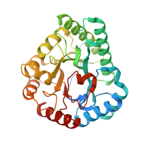
    d-Allulose has potential as a low-calorie sweetener which can suppress fat accumulation. Several enzymes capable of d-allulose production have been isolated, including d-tagatose 3-epimerases. Here, we report the isolation of a novel protein from Methylomonas sp. expected to be a putative enzyme based on sequence similarity to ketose 3-epimerase. The synthesized gene encoding the deduced ketose 3-epimerase was expressed as a recombinant enzyme in Escherichia coli, and it exhibited the highest enzymatic activity toward l-ribulose, followed by d-ribulose and d-allulose. The X-ray structure analysis of l-ribulose 3-epimerase from Methylomonas sp. (MetLRE) revealed a homodimeric enzyme, the first reported structure of dimeric l-ribulose 3-epimerase. The monomeric structure of MetLRE is similar to that of homotetrameric l-ribulose 3-epimerases, but the short C-terminal α-helix of MetLRE is unique and different from those of known l-ribulose 3 epimerases. The length of the C-terminal α-helix was thought to be involved in tetramerization and increasing stability; however, the addition of residues to MetLRE at the C terminus did not lead to tetramer formation. MetLRE is the first dimeric l-ribulose 3-epimerase identified to exhibit high relative activity toward d-allulose.


  • Organizational Affiliation

    Life Science Research Center and Faculty of Medicine, Kagawa University, Kita, Japan.


Macromolecules
Find similar proteins by:  (by identity cutoff)  |  3D Structure
Entity ID: 1
MoleculeChains Sequence LengthOrganismDetailsImage
Epimerase
A, B, C, D, E
A, B, C, D, E, F
307Methylomonas sp. DH-1Mutation(s): 0 
Gene Names: AYM39_05640
UniProt
Find proteins for A0A172U6X0 (Methylomonas sp. (strain DH-1))
Explore A0A172U6X0 
Go to UniProtKB:  A0A172U6X0
Entity Groups  
Sequence Clusters30% Identity50% Identity70% Identity90% Identity95% Identity100% Identity
UniProt GroupA0A172U6X0
Sequence Annotations
Expand
  • Reference Sequence
Small Molecules
Ligands 3 Unique
IDChains Name / Formula / InChI Key2D Diagram3D Interactions
EPE
Query on EPE

Download Ideal Coordinates CCD File 
H [auth A]
K [auth B]
N [auth C]
Q [auth D]
T [auth E]
H [auth A],
K [auth B],
N [auth C],
Q [auth D],
T [auth E],
W [auth F]
4-(2-HYDROXYETHYL)-1-PIPERAZINE ETHANESULFONIC ACID
C8 H18 N2 O4 S
JKMHFZQWWAIEOD-UHFFFAOYSA-N
PSJ
Query on PSJ

Download Ideal Coordinates CCD File 
I [auth A]
L [auth B]
O [auth C]
R [auth D]
U [auth E]
I [auth A],
L [auth B],
O [auth C],
R [auth D],
U [auth E],
X [auth F]
D-psicose
C6 H12 O6
BJHIKXHVCXFQLS-PUFIMZNGSA-N
MN
Query on MN

Download Ideal Coordinates CCD File 
G [auth A]
J [auth B]
M [auth C]
P [auth D]
S [auth E]
G [auth A],
J [auth B],
M [auth C],
P [auth D],
S [auth E],
V [auth F]
MANGANESE (II) ION
Mn
WAEMQWOKJMHJLA-UHFFFAOYSA-N
Experimental Data & Validation

Experimental Data

  • Method: X-RAY DIFFRACTION
  • Resolution: 2.05 Å
  • R-Value Free: 0.241 
  • R-Value Work: 0.162 
  • R-Value Observed: 0.166 
  • Space Group: P 1
Unit Cell:
Length ( Å )Angle ( ˚ )
a = 51.02α = 99.3
b = 81.05β = 101.5
c = 106.13γ = 87.24
Software Package:
Software NamePurpose
XDSdata reduction
XSCALEdata scaling
REFMACrefinement
PDB_EXTRACTdata extraction
MOLREPphasing

Structure Validation

View Full Validation Report


Ligand Structure Quality Assessment 


Entry History 

Deposition Data

Revision History  (Full details and data files)

  • Version 1.0: 2021-04-21
    Type: Initial release
  • Version 1.1: 2021-06-09
    Changes: Database references
  • Version 1.2: 2023-11-29
    Changes: Data collection, Database references, Refinement description