7ANQ

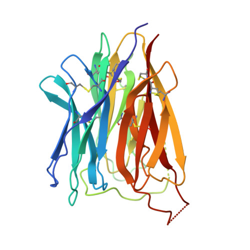
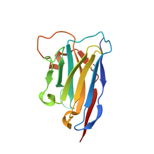
Complete PCSK9 C-ter domain in complex with VHH P1.40


Experimental Data Snapshot

  • Method: X-RAY DIFFRACTION
  • Resolution: 2.20 Å
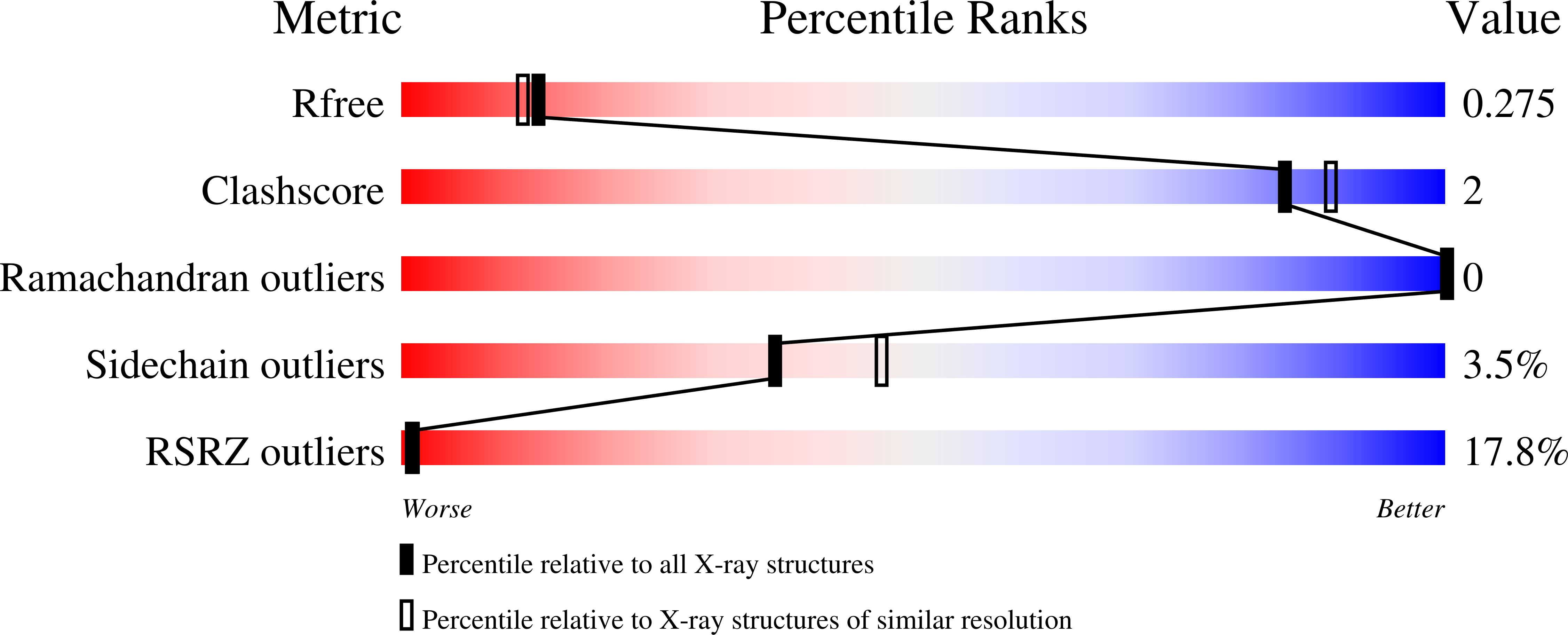
  • R-Value Free: 0.256 
  • R-Value Work: 0.213 
  • R-Value Observed: 0.215 

wwPDB Validation   3D Report Full Report


Ligand Structure Quality Assessment 


This is version 1.2 of the entry. See complete history


Literature

Molecular interactions of PCSK9 with an inhibitory nanobody, CAP1 and HLA-C: Functional regulation of LDLR levels.

Fruchart Gaillard, C.Ouadda, A.B.D.Ciccone, L.Girard, E.Mikaeeli, S.Evagelidis, A.Le Devehat, M.Susan-Resiga, D.Lajeunesse, E.C.Nozach, H.Ramos, O.H.P.Thureau, A.Legrand, P.Prat, A.Dive, V.Seidah, N.G.

(2022) Mol Metab 67: 101662-101662

  • DOI: https://doi.org/10.1016/j.molmet.2022.101662
  • Primary Citation of Related Structures:  
    7ANQ

  • PubMed Abstract: 

    The liver-derived circulating PCSK9 enhances the degradation of the LDL receptor (LDLR) in endosomes/lysosomes. PCSK9 inhibition or silencing is presently used in clinics worldwide to reduce LDL-cholesterol, resulting in lower incidence of cardiovascular disease and possibly cancer/metastasis. The mechanism by which the PCSK9-LDLR complex is sorted to degradation compartments is not fully understood. We previously suggested that out of the three M1, M2 and M3 subdomains of the C-terminal Cys/His-rich-domain (CHRD) of PCSK9, only M2 is critical for the activity of extracellular of PCSK9 on cell surface LDLR. This likely implicates the binding of M2 to an unknown membrane-associated "protein X" that would escort the complex to endosomes/lysosomes for degradation. We reported that a nanobody P1.40 binds the M1 and M3 domains of the CHRD and inhibits the function of PCSK9. It was also reported that the cytosolic adenylyl cyclase-associated protein 1 (CAP1) could bind M1 and M3 subdomains and enhance the activity of PCSK9. In this study, we determined the 3-dimensional structure of the CHRD-P1.40 complex to understand the intricate interplay between P1.40, CAP1 and PCSK9 and how they regulate LDLR degradation.


  • Organizational Affiliation

    Université Paris-Saclay, CEA, INRAE, Département Médicaments et Technologies pour la Santé (DMTS), SIMoS, 91191 Gif-sur-Yvette, France.


Macromolecules
Find similar proteins by:  (by identity cutoff)  |  3D Structure
Entity ID: 1
MoleculeChains Sequence LengthOrganismDetailsImage
Proprotein convertase subtilisin/kexin type 9231Homo sapiensMutation(s): 0 
Gene Names: PCSK9NARC1PSEC0052
EC: 3.4.21
UniProt & NIH Common Fund Data Resources
Find proteins for Q8NBP7 (Homo sapiens)
Explore Q8NBP7 
Go to UniProtKB:  Q8NBP7
PHAROS:  Q8NBP7
GTEx:  ENSG00000169174 
Entity Groups  
Sequence Clusters30% Identity50% Identity70% Identity90% Identity95% Identity100% Identity
UniProt GroupQ8NBP7
Sequence Annotations
Expand
  • Reference Sequence
Find similar proteins by:  (by identity cutoff)  |  3D Structure
Entity ID: 2
MoleculeChains Sequence LengthOrganismDetailsImage
VHH P1.40 minibody anti-Cter PCSK9127Lama glamaMutation(s): 0 
Entity Groups  
Sequence Clusters30% Identity50% Identity70% Identity90% Identity95% Identity100% Identity
Sequence Annotations
Expand
  • Reference Sequence
Oligosaccharides

Help

Entity ID: 3
MoleculeChains Length2D Diagram Glycosylation3D Interactions
2-acetamido-2-deoxy-beta-D-glucopyranose-(1-4)-[alpha-L-fucopyranose-(1-6)]2-acetamido-2-deoxy-beta-D-glucopyranose
C
3N-Glycosylation
Glycosylation Resources
GlyTouCan:  G21290RB
GlyCosmos:  G21290RB
GlyGen:  G21290RB
Small Molecules
Ligands 1 Unique
IDChains Name / Formula / InChI Key2D Diagram3D Interactions
SO4 (Subject of Investigation/LOI)
Query on SO4

Download Ideal Coordinates CCD File 
D [auth A]SULFATE ION
O4 S
QAOWNCQODCNURD-UHFFFAOYSA-L
Experimental Data & Validation

Experimental Data

  • Method: X-RAY DIFFRACTION
  • Resolution: 2.20 Å
  • R-Value Free: 0.256 
  • R-Value Work: 0.213 
  • R-Value Observed: 0.215 
  • Space Group: P 41 21 2
Unit Cell:
Length ( Å )Angle ( ˚ )
a = 52.43α = 90
b = 52.43β = 90
c = 263.41γ = 90
Software Package:
Software NamePurpose
BUSTERrefinement
PDB_EXTRACTdata extraction
XDSdata reduction
XSCALEdata scaling
MOLREPmodel building
PHASERphasing

Structure Validation

View Full Validation Report


Ligand Structure Quality Assessment 


Entry History 

Deposition Data

Revision History  (Full details and data files)

  • Version 1.0: 2021-10-20
    Type: Initial release
  • Version 1.1: 2023-01-25
    Changes: Database references
  • Version 1.2: 2024-02-07
    Changes: Data collection, Refinement description