7AH5

Crystal structure of indoleamine 2,3-dioxygenase 1 (IDO1) in complex with ferric heme and MMG-0706


Experimental Data Snapshot

  • Method: X-RAY DIFFRACTION
  • Resolution: 2.90 Å
  • R-Value Free: 0.292 
  • R-Value Work: 0.231 
  • R-Value Observed: 0.237 

wwPDB Validation   3D Report Full Report


Ligand Structure Quality Assessment 


This is version 1.3 of the entry. See complete history


Literature

Azole-Based Indoleamine 2,3-Dioxygenase 1 (IDO1) Inhibitors.

Rohrig, U.F.Majjigapu, S.R.Reynaud, A.Pojer, F.Dilek, N.Reichenbach, P.Ascencao, K.Irving, M.Coukos, G.Vogel, P.Michielin, O.Zoete, V.

(2021) J Med Chem 64: 2205-2227

  • DOI: https://doi.org/10.1021/acs.jmedchem.0c01968
  • Primary Citation of Related Structures:  
    7AH4, 7AH5, 7AH6

  • PubMed Abstract: 

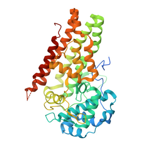
    The heme enzyme indoleamine 2,3-dioxygenase 1 (IDO1) plays an essential role in immunity, neuronal function, and aging through catalysis of the rate-limiting step in the kynurenine pathway of tryptophan metabolism. Many IDO1 inhibitors with different chemotypes have been developed, mainly targeted for use in anti-cancer immunotherapy. Lead optimization of direct heme iron-binding inhibitors has proven difficult due to the remarkable selectivity and sensitivity of the heme-ligand interactions. Here, we present experimental data for a set of closely related small azole compounds with more than 4 orders of magnitude differences in their inhibitory activities, ranging from millimolar to nanomolar levels. We investigate and rationalize their activities based on structural data, molecular dynamics simulations, and density functional theory calculations. Our results not only expand the presently known four confirmed chemotypes of sub-micromolar heme binding IDO1 inhibitors by two additional scaffolds but also provide a model to predict the activities of novel scaffolds.


  • Organizational Affiliation

    Molecular Modeling Group, SIB Swiss Institute of Bioinformatics, 1015 Lausanne, Switzerland.


Macromolecules
Find similar proteins by:  (by identity cutoff)  |  3D Structure
Entity ID: 1
MoleculeChains Sequence LengthOrganismDetailsImage
Indoleamine 2,3-dioxygenase 1
A, B
423Homo sapiensMutation(s): 0 
Gene Names: IDO1IDOINDO
EC: 1.13.11.52
UniProt & NIH Common Fund Data Resources
Find proteins for P14902 (Homo sapiens)
Explore P14902 
Go to UniProtKB:  P14902
PHAROS:  P14902
GTEx:  ENSG00000131203 
Entity Groups  
Sequence Clusters30% Identity50% Identity70% Identity90% Identity95% Identity100% Identity
UniProt GroupP14902
Sequence Annotations
Expand
  • Reference Sequence
Experimental Data & Validation

Experimental Data

  • Method: X-RAY DIFFRACTION
  • Resolution: 2.90 Å
  • R-Value Free: 0.292 
  • R-Value Work: 0.231 
  • R-Value Observed: 0.237 
  • Space Group: P 21 21 21
Unit Cell:
Length ( Å )Angle ( ˚ )
a = 84.988α = 90
b = 93.106β = 90
c = 130.36γ = 90
Software Package:
Software NamePurpose
PHENIXrefinement
PDB_EXTRACTdata extraction
XDSdata reduction
XDSdata scaling
PHASERphasing

Structure Validation

View Full Validation Report


Ligand Structure Quality Assessment 


Entry History 

Deposition Data

Revision History  (Full details and data files)

  • Version 1.0: 2021-02-17
    Type: Initial release
  • Version 1.1: 2021-02-24
    Changes: Database references
  • Version 1.2: 2021-03-10
    Changes: Database references
  • Version 1.3: 2024-01-31
    Changes: Data collection, Database references, Refinement description