


Crystal structure of a Putative structural protein from Klebsiella pneumoniae
Liu, L., Lovell, S., Battaile, K.P., Tillery, L., Shek, R., Craig, J.K., Barrett, L.K., Van Voorhis, W.C.To be published.
Experimental Data Snapshot
Starting Model: experimental
View more details
wwPDB Validation 3D Report Full Report

Entity ID: 1 | |||||
|---|---|---|---|---|---|
| Molecule | Chains | Sequence Length | Organism | Details | Image |
| Putative structural protein | 172 | Klebsiella pneumoniae subsp. pneumoniae HS11286 | Mutation(s): 0 Gene Names: KPHS_20300 | 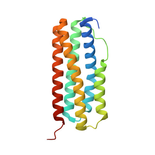 | |
UniProt | |||||
Find proteins for A0A0H3GMY9 (Klebsiella pneumoniae subsp. pneumoniae (strain HS11286)) Explore A0A0H3GMY9 Go to UniProtKB: A0A0H3GMY9 | |||||
Entity Groups | |||||
| Sequence Clusters | 30% Identity50% Identity70% Identity90% Identity95% Identity100% Identity | ||||
| UniProt Group | A0A0H3GMY9 | ||||
Sequence AnnotationsExpand | |||||
| |||||
| Ligands 1 Unique | |||||
|---|---|---|---|---|---|
| ID | Chains | Name / Formula / InChI Key | 2D Diagram | 3D Interactions | |
| ZN (Subject of Investigation/LOI) Query on ZN | C [auth A], D [auth A], E [auth B], F [auth B] | ZINC ION Zn PTFCDOFLOPIGGS-UHFFFAOYSA-N |  | ||
| Length ( Å ) | Angle ( ˚ ) |
|---|---|
| a = 62.227 | α = 90 |
| b = 104.652 | β = 90 |
| c = 45.889 | γ = 90 |
| Software Name | Purpose |
|---|---|
| XDS | data reduction |
| Aimless | data scaling |
| MOLREP | phasing |
| PHENIX | refinement |
| PDB_EXTRACT | data extraction |
| Funding Organization | Location | Grant Number |
|---|---|---|
| National Institutes of Health/National Institute Of Allergy and Infectious Diseases (NIH/NIAID) | United States | HHSN272201700059C |
RCSB PDB (citation) is hosted by
RCSB PDB is a member of the

