


Crystal Structure of sulfurtransferase (DsrC family protein) from Acinetobacter baumannii
Calhoun, B.M., Bolejack, M.J., Lorimer, D.D., Horanyi, P.S., Edwards, T.E.To be published.
Experimental Data Snapshot
wwPDB Validation 3D Report Full Report

Entity ID: 1 | |||||
|---|---|---|---|---|---|
| Molecule | Chains | Sequence Length | Organism | Details | Image |
| Sulfurtransferase | 111 | Acinetobacter baumannii | Mutation(s): 0 Gene Names: tusE EC: 2.8.1 | 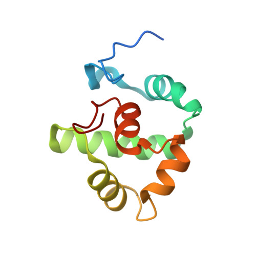 | |
UniProt | |||||
Find proteins for V5VCI8 (Acinetobacter baumannii) Explore V5VCI8 Go to UniProtKB: V5VCI8 | |||||
Entity Groups | |||||
| Sequence Clusters | 30% Identity50% Identity70% Identity90% Identity95% Identity100% Identity | ||||
| UniProt Group | V5VCI8 | ||||
Sequence AnnotationsExpand | |||||
| |||||
| Length ( Å ) | Angle ( ˚ ) |
|---|---|
| a = 32.5 | α = 90 |
| b = 46.84 | β = 108.02 |
| c = 39.48 | γ = 90 |
| Software Name | Purpose |
|---|---|
| XDS | data reduction |
| XSCALE | data scaling |
| PHENIX | refinement |
| PDB_EXTRACT | data extraction |
| MR-Rosetta | phasing |
| Funding Organization | Location | Grant Number |
|---|---|---|
| National Institutes of Health/National Institute Of Allergy and Infectious Diseases (NIH/NIAID) | United States | -- |
RCSB PDB (citation) is hosted by
RCSB PDB is a member of the

