The structure and function of modular Escherichia coli O157:H7 bacteriophage FTBEc1 endolysin, LysT84: defining a new endolysin catalytic subfamily.
Love, M.J., Coombes, D., Ismail, S., Billington, C., Dobson, R.C.J.(2022) Biochem J 479: 207-223
- PubMed: 34935873
- DOI: https://doi.org/10.1042/BCJ20210701
- Primary Citation of Related Structures:
7RUM - PubMed Abstract:
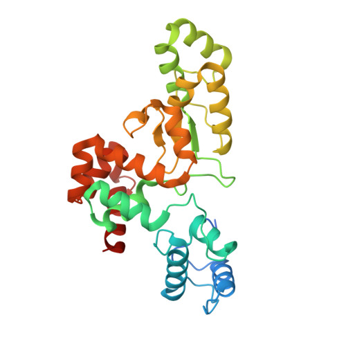
Bacteriophage endolysins degrade peptidoglycan and have been identified as antibacterial candidates to combat antimicrobial resistance. Considering the catalytic and structural diversity of endolysins, there is a paucity of structural data to inform how these enzymes work at the molecular level - key data that is needed to realize the potential of endolysin-based antibacterial agents. Here, we determine the atomic structure and define the enzymatic function of Escherichia coli O157:H7 phage FTEBc1 endolysin, LysT84. Bioinformatic analysis reveals that LysT84 is a modular endolysin, which is unusual for Gram-negative endolysins, comprising a peptidoglycan binding domain and an enzymatic domain. The crystal structure of LysT84 (2.99 Å) revealed a mostly α-helical protein with two domains connected by a linker region but packed together. LysT84 was determined to be a monomer in solution using analytical ultracentrifugation. Small-angle X-ray scattering data revealed that LysT84 is a flexible protein but does not have the expected bimodal P(r) function of a multidomain protein, suggesting that the domains of LysT84 pack closely creating a globular protein as seen in the crystal structure. Structural analysis reveals two key glutamate residues positioned on either side of the active site cavity; mutagenesis demonstrating these residues are critical for peptidoglycan degradation. Molecular dynamic simulations suggest that the enzymatically active domain is dynamic, allowing the appropriate positioning of these catalytic residues for hydrolysis of the β(1-4) bond. Overall, our study defines the structural basis for peptidoglycan degradation by LysT84 which supports rational engineering of related endolysins into effective antibacterial agents.
Organizational Affiliation:
Biomolecular Interaction Centre and School of Biological Sciences, University of Canterbury, Christchurch, New Zealand.