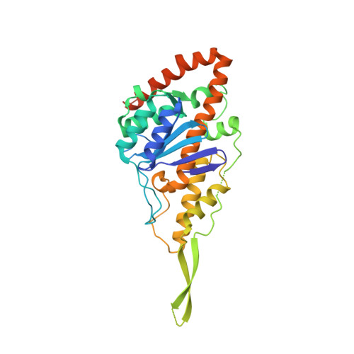
Crystal structure of DNA polymerase I from Thermus phage G20c.
Ahlqvist, J., Linares-Pasten, J.A., Jasilionis, A., Welin, M., Hakansson, M., Svensson, L.A., Wang, L., Watzlawick, H., Aevarsson, A., Fridjonsson, O.H., Hreggvidsson, G.O., Ketelsen Striberny, B., Glomsaker, E., Lanes, O., Al-Karadaghi, S., Nordberg Karlsson, E.(2022) Acta Crystallogr D Struct Biol 78: 1384-1398
- PubMed: 36322421
- DOI: https://doi.org/10.1107/S2059798322009895
- Primary Citation of Related Structures:
7R0K, 7R0T - PubMed Abstract:
This study describes the structure of DNA polymerase I from Thermus phage G20c, termed PolI_G20c. This is the first structure of a DNA polymerase originating from a group of related thermophilic bacteriophages infecting Thermus thermophilus, including phages G20c, TSP4, P74-26, P23-45 and phiFA and the novel phage Tth15-6. Sequence and structural analysis of PolI_G20c revealed a 3'-5' exonuclease domain and a DNA polymerase domain, and activity screening confirmed that both domains were functional. No functional 5'-3' exonuclease domain was present. Structural analysis also revealed a novel specific structure motif, here termed SβαR, that was not previously identified in any polymerase belonging to the DNA polymerases I (or the DNA polymerase A family). The SβαR motif did not show any homology to the sequences or structures of known DNA polymerases. The exception was the sequence conservation of the residues in this motif in putative DNA polymerases encoded in the genomes of a group of thermophilic phages related to Thermus phage G20c. The structure of PolI_G20c was determined with the aid of another structure that was determined in parallel and was used as a model for molecular replacement. This other structure was of a 3'-5' exonuclease termed ExnV1. The cloned and expressed gene encoding ExnV1 was isolated from a thermophilic virus metagenome that was collected from several hot springs in Iceland. The structure of ExnV1, which contains the novel SβαR motif, was first determined to 2.19 Å resolution. With these data at hand, the structure of PolI_G20c was determined to 2.97 Å resolution. The structures of PolI_G20c and ExnV1 are most similar to those of the Klenow fragment of DNA polymerase I (PDB entry 2kzz) from Escherichia coli, DNA polymerase I from Geobacillus stearothermophilus (PDB entry 1knc) and Taq polymerase (PDB entry 1bgx) from Thermus aquaticus.
Organizational Affiliation:
Division of Biotechnology, Department of Chemistry, Lund University, PO Box 124, 221 00 Lund, Sweden.


