7PH1

Trypsin in complex with BPTI mutant (2S)-2-amino-4-monofluorobutanoic acid


Experimental Data Snapshot

  • Method: X-RAY DIFFRACTION
  • Resolution: 1.18 Å
  • R-Value Free: 0.166 
  • R-Value Work: 0.149 
  • R-Value Observed: 0.149 

wwPDB Validation   3D Report Full Report


This is version 1.4 of the entry. See complete history


Literature

Water Network in the Binding Pocket of Fluorinated BPTI-Trypsin Complexes─Insights from Simulation and Experiment.

Wehrhan, L.Leppkes, J.Dimos, N.Loll, B.Koksch, B.Keller, B.G.

(2022) J Phys Chem B 126: 9985-9999

  • DOI: https://doi.org/10.1021/acs.jpcb.2c05496
  • Primary Citation of Related Structures:  
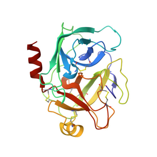
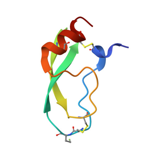
    7PH1

  • PubMed Abstract: 

    Structural waters in the S1 binding pocket of β-trypsin are critical for the stabilization of the complex of β-trypsin with its inhibitor bovine pancreatic trypsin inhibitor (BPTI). The inhibitor strength of BPTI can be modulated by replacing the critical lysine residue at the P1 position by non-natural amino acids. We study BPTI variants in which the critical Lys15 in BPTI has been replaced by α-aminobutyric acid (Abu) and its fluorinated derivatives monofluoroethylglycine (MfeGly), difluoroethylglycine (DfeGly), and trifluoroethylglycine (TfeGly). We investigate the hypothesis that additional water molecules in the binding pocket can form specific noncovalent interactions with the fluorinated side chains and thereby act as an extension of the inhibitors. We report potentials of mean force (PMF) of the unbinding process for all four complexes and enzyme activity inhibition assays. Additionally, we report the protein crystal structure of the Lys15MfeGly-BPTI-β-trypsin complex (pdb: 7PH1). Both experimental and computational data show a stepwise increase in inhibitor strength with increasing fluorination of the Abu side chain. The PMF additionally shows a minimum for the encounter complex and an intermediate state just before the bound state. In the bound state, the computational analysis of the structure and dynamics of the water molecules in the S1 pocket shows a highly dynamic network of water molecules that does not indicate a rigidification or stabilizing trend in regard to energetic properties that could explain the increase in inhibitor strength. The analysis of the energy and the entropy of the water molecules in the S1 binding pocket using grid inhomogeneous solvation theory confirms this result. Overall, fluorination systematically changes the binding affinity, but the effect cannot be explained by a persistent water network in the binding pocket. Other effects, such as the hydrophobicity of fluorinated amino acids and the stability of the encounter complex as well as the additional minimum in the potential of mean force in the bound state, likely influence the affinity more directly.


  • Organizational Affiliation

    Department of Biology, Chemistry, and Pharmacy, Freie Universität Berlin, Institute of Chemistry and Biochemistry, Arnimallee 22, Berlin14195, Germany.


Macromolecules
Find similar proteins by:  (by identity cutoff)  |  3D Structure
Entity ID: 1
MoleculeChains Sequence LengthOrganismDetailsImage
Cationic trypsinA [auth E]223Bos taurusMutation(s): 0 
EC: 3.4.21.4
UniProt
Find proteins for P00760 (Bos taurus)
Explore P00760 
Go to UniProtKB:  P00760
Entity Groups  
Sequence Clusters30% Identity50% Identity70% Identity90% Identity95% Identity100% Identity
UniProt GroupP00760
Sequence Annotations
Expand
  • Reference Sequence
Find similar proteins by:  (by identity cutoff)  |  3D Structure
Entity ID: 2
MoleculeChains Sequence LengthOrganismDetailsImage
Pancreatic trypsin inhibitorB [auth I]57Bos taurusMutation(s): 0 
UniProt
Find proteins for P00974 (Bos taurus)
Explore P00974 
Go to UniProtKB:  P00974
Entity Groups  
Sequence Clusters30% Identity50% Identity70% Identity90% Identity95% Identity100% Identity
UniProt GroupP00974
Sequence Annotations
Expand
  • Reference Sequence
Small Molecules
Ligands 3 Unique
IDChains Name / Formula / InChI Key2D Diagram3D Interactions
SO4
Query on SO4

Download Ideal Coordinates CCD File 
C [auth E]
J [auth E]
K [auth E]
L [auth E]
M [auth E]
C [auth E],
J [auth E],
K [auth E],
L [auth E],
M [auth E],
O [auth E],
P [auth E],
Q [auth E],
R [auth E],
U [auth I],
V [auth I],
W [auth I],
X [auth I]
SULFATE ION
O4 S
QAOWNCQODCNURD-UHFFFAOYSA-L
GOL
Query on GOL

Download Ideal Coordinates CCD File 
D [auth E]
E
F [auth E]
G [auth E]
H [auth E]
D [auth E],
E,
F [auth E],
G [auth E],
H [auth E],
I [auth E],
S [auth I],
T [auth I],
Y [auth I]
GLYCEROL
C3 H8 O3
PEDCQBHIVMGVHV-UHFFFAOYSA-N
CA
Query on CA

Download Ideal Coordinates CCD File 
N [auth E]CALCIUM ION
Ca
BHPQYMZQTOCNFJ-UHFFFAOYSA-N
Modified Residues  1 Unique
IDChains TypeFormula2D DiagramParent
7OZ
Query on 7OZ
B [auth I]L-PEPTIDE LINKINGC4 H8 F N O2ALA
Experimental Data & Validation

Experimental Data

Unit Cell:
Length ( Å )Angle ( ˚ )
a = 74.975α = 90
b = 81.289β = 90
c = 124.248γ = 90
Software Package:
Software NamePurpose
PHENIXrefinement
PDB_EXTRACTdata extraction
XDSdata reduction
XSCALEdata scaling
PHASERphasing

Structure Validation

View Full Validation Report


Entry History & Funding Information

Deposition Data


Funding OrganizationLocationGrant Number
German Research Foundation (DFG)GermanySfB 1349 / 387284271

Revision History  (Full details and data files)

  • Version 1.0: 2022-03-30
    Type: Initial release
  • Version 1.1: 2022-10-12
    Changes: Database references
  • Version 1.2: 2022-11-30
    Changes: Database references
  • Version 1.3: 2022-12-21
    Changes: Database references
  • Version 1.4: 2024-01-31
    Changes: Data collection, Refinement description