7N52

Structure of a bacterial gasdermin from Runella zeae


Experimental Data Snapshot

  • Method: X-RAY DIFFRACTION
  • Resolution: 2.90 Å
  • R-Value Free: 0.280 
  • R-Value Work: 0.237 
  • R-Value Observed: 0.241 

wwPDB Validation   3D Report Full Report


This is version 1.1 of the entry. See complete history


Literature

Bacterial gasdermins reveal an ancient mechanism of cell death.

Johnson, A.G.Wein, T.Mayer, M.L.Duncan-Lowey, B.Yirmiya, E.Oppenheimer-Shaanan, Y.Amitai, G.Sorek, R.Kranzusch, P.J.

(2022) Science 375: 221-225

  • DOI: https://doi.org/10.1126/science.abj8432
  • Primary Citation of Related Structures:  
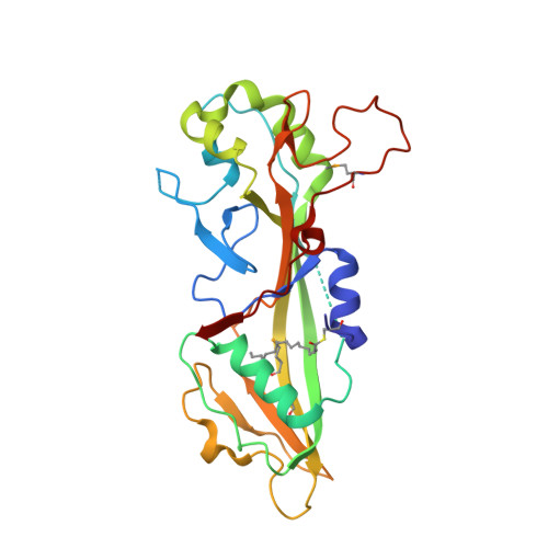
    7N50, 7N51, 7N52

  • PubMed Abstract: 

    Gasdermin proteins form large membrane pores in human cells that release immune cytokines and induce lytic cell death. Gasdermin pore formation is triggered by caspase-mediated cleavage during inflammasome signaling and is critical for defense against pathogens and cancer. We discovered gasdermin homologs encoded in bacteria that defended against phages and executed cell death. Structures of bacterial gasdermins revealed a conserved pore-forming domain that was stabilized in the inactive state with a buried lipid modification. Bacterial gasdermins were activated by dedicated caspase-like proteases that catalyzed site-specific cleavage and the removal of an inhibitory C-terminal peptide. Release of autoinhibition induced the assembly of large and heterogeneous pores that disrupted membrane integrity. Thus, pyroptosis is an ancient form of regulated cell death shared between bacteria and animals.


  • Organizational Affiliation

    Department of Microbiology, Harvard Medical School, Boston, MA 02115, USA.


Macromolecules
Find similar proteins by:  (by identity cutoff)  |  3D Structure
Entity ID: 1
MoleculeChains Sequence LengthOrganismDetailsImage
Gasdermin
A, B, C, D
268Runella zeaeMutation(s): 0 
Entity Groups  
Sequence Clusters30% Identity50% Identity70% Identity90% Identity95% Identity100% Identity
Sequence Annotations
Expand
  • Reference Sequence
Small Molecules
Modified Residues  2 Unique
IDChains TypeFormula2D DiagramParent
MSE
Query on MSE
A, B, C, D
L-PEPTIDE LINKINGC5 H11 N O2 SeMET
P1L
Query on P1L
A, B, C, D
L-PEPTIDE LINKINGC19 H37 N O3 SCYS
Experimental Data & Validation

Experimental Data

  • Method: X-RAY DIFFRACTION
  • Resolution: 2.90 Å
  • R-Value Free: 0.280 
  • R-Value Work: 0.237 
  • R-Value Observed: 0.241 
  • Space Group: P 1 21 1
Unit Cell:
Length ( Å )Angle ( ˚ )
a = 69.29α = 90
b = 103.852β = 111.544
c = 78.992γ = 90
Software Package:
Software NamePurpose
PHENIXrefinement
XDSdata reduction
Aimlessdata scaling
PHENIXphasing

Structure Validation

View Full Validation Report


Entry History & Funding Information

Deposition Data


Funding OrganizationLocationGrant Number
National Institutes of Health/National Cancer Institute (NIH/NCI)United StatesT32CA207021

Revision History  (Full details and data files)

  • Version 1.0: 2021-06-23
    Type: Initial release
  • Version 1.1: 2022-01-26
    Changes: Database references