7LR8

Crystal structure of GH5_18-E153A from Streptomyces cattleya in complex with Manb1-4GlcNAc


Experimental Data Snapshot

  • Method: X-RAY DIFFRACTION
  • Resolution: 1.60 Å
  • R-Value Free: 0.191 
  • R-Value Work: 0.162 
  • R-Value Observed: 0.163 

wwPDB Validation   3D Report Full Report


This is version 1.2 of the entry. See complete history


Literature

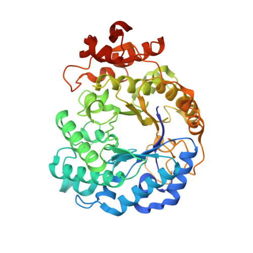
N-Glycan Degradation Pathways in Gut- and Soil-Dwelling Actinobacteria Share Common Core Genes.

Higgins, M.A.Tegl, G.MacDonald, S.S.Arnal, G.Brumer, H.Withers, S.G.Ryan, K.S.

(2021) ACS Chem Biol 16: 701-711

  • DOI: https://doi.org/10.1021/acschembio.0c00995
  • Primary Citation of Related Structures:  
    7LQX, 7LR1, 7LR2, 7LR6, 7LR7, 7LR8

  • PubMed Abstract: 

    N-Glycosylation is a fundamental protein modification found in both eukaryotes and archaea. Despite lacking N-glycans, many commensal and pathogenic bacteria have developed mechanisms to degrade these isoforms for a variety of functions, including nutrient acquisition and evasion of the immune system. Although much is known about many of the enzymes responsible for N-glycan degradation, the enzymes involved in cleaving the N-glycan core have only recently been discovered. Thus, some of the structural details have yet to be characterized, and little is known about their full distribution among bacterial strains and specifically within potential Gram-positive polysaccharide utilization loci. Here, we report crystal structures for Family 5, Subfamily 18 (GH5_18) glycoside hydrolases from the gut bacterium Bifidobacterium longum (BlGH5_18) and the soil bacterium Streptomyces cattleya (ScGH5_18), which hydrolyze the core Manβ1-4GlcNAc disaccharide. Structures of these enzymes in complex with Manβ1-4GlcNAc reveal a more complete picture of the -1 subsite. They also show that a C-terminal active site cap present in BlGH5_18 is absent in ScGH5_18. Although this C-terminal cap is not widely distributed throughout the GH5_18 family, it is important for full enzyme activity. In addition, we show that GH5_18 enzymes are found in Gram-positive polysaccharide utilization loci that share common genes, likely dedicated to importing and degrading N-glycan core structures.


Macromolecules
Find similar proteins by:  (by identity cutoff)  |  3D Structure
Entity ID: 1
MoleculeChains Sequence LengthOrganismDetailsImage
Uncharacterized protein ScGH5_18
A, B
433Streptantibioticus cattleyicolor NRRL 8057 = DSM 46488Mutation(s): 1 
Gene Names: SCATT_p07120
UniProt
Find proteins for F8JJ04 (Streptantibioticus cattleyicolor (strain ATCC 35852 / DSM 46488 / JCM 4925 / NBRC 14057 / NRRL 8057))
Explore F8JJ04 
Go to UniProtKB:  F8JJ04
Entity Groups  
Sequence Clusters30% Identity50% Identity70% Identity90% Identity95% Identity100% Identity
UniProt GroupF8JJ04
Sequence Annotations
Expand
  • Reference Sequence
Oligosaccharides

Help

Entity ID: 2
MoleculeChains Length2D Diagram Glycosylation3D Interactions
beta-D-mannopyranose-(1-4)-2-acetamido-2-deoxy-beta-D-glucopyranoseC [auth E],
D [auth C]
2N/A
Glycosylation Resources
GlyTouCan:  G90333CG
GlyCosmos:  G90333CG
GlyGen:  G90333CG
Experimental Data & Validation

Experimental Data

  • Method: X-RAY DIFFRACTION
  • Resolution: 1.60 Å
  • R-Value Free: 0.191 
  • R-Value Work: 0.162 
  • R-Value Observed: 0.163 
  • Space Group: C 1 2 1
Unit Cell:
Length ( Å )Angle ( ˚ )
a = 152.869α = 90
b = 76.455β = 117.24
c = 79.92γ = 90
Software Package:
Software NamePurpose
PHENIXrefinement
XDSdata reduction
Aimlessdata scaling
PHASERphasing

Structure Validation

View Full Validation Report


Entry History & Funding Information

Deposition Data


Funding OrganizationLocationGrant Number
Canadian Institutes of Health Research (CIHR)Canada--

Revision History  (Full details and data files)

  • Version 1.0: 2021-04-07
    Type: Initial release
  • Version 1.1: 2021-04-28
    Changes: Database references
  • Version 1.2: 2023-10-18
    Changes: Data collection, Database references, Refinement description