7LD9

Structure of human GGT1 in complex with ABBA


Experimental Data Snapshot

  • Method: X-RAY DIFFRACTION
  • Resolution: 1.42 Å
  • R-Value Free: 0.134 
  • R-Value Work: 0.107 
  • R-Value Observed: 0.108 

wwPDB Validation   3D Report Full Report


Ligand Structure Quality Assessment 


This is version 1.2 of the entry. See complete history


Literature

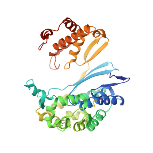
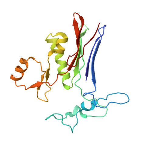
Design and evaluation of novel analogs of 2-amino-4-boronobutanoic acid (ABBA) as inhibitors of human gamma-glutamyl transpeptidase.

Nguyen, L.Schultz, D.C.Terzyan, S.S.Rezaei, M.Songb, J.Li, C.You, Y.Hanigan, M.H.

(2022) Bioorg Med Chem 73: 116986-116986

  • DOI: https://doi.org/10.1016/j.bmc.2022.116986
  • Primary Citation of Related Structures:  
    7LBC, 7LD9

  • PubMed Abstract: 

    Inhibitors of gamma-glutamyl transpeptidase (GGT1, aka gamma-glutamyl transferase) are needed for the treatment of cancer, cardiovascular illness and other diseases. Compounds that inhibit GGT1 have been evaluated in the clinic, but no inhibitor has successfully demonstrated specific and systemic GGT1 inhibition. All have severe side effects. L-2-amino-4‑boronobutanoic acid (l-ABBA), a glutamate analog, is the most potent GGT1 inhibitor in vitro. In this study, we have solved the crystal structure of human GGT1 (hGGT1) with ABBA bound in the active site. The structure was interrogated to identify interactions between the enzyme and the inhibitor. Based on these data, a series of novel ABBA analogs were designed and synthesized. Their inhibitory activity against the hydrolysis and transpeptidation activities of hGGT1 were determined. The lead compounds were crystalized with hGGT1 and the structures solved. The kinetic data and structures of the complexes provide new insights into the critical role of protein structure dynamics in developing compounds for inhibition of hGGT1.


  • Organizational Affiliation

    Department of Pharmaceutical Sciences, The State University of New York at Buffalo, Buffalo, NY 14214, United States.


Macromolecules
Find similar proteins by:  (by identity cutoff)  |  3D Structure
Entity ID: 1
MoleculeChains Sequence LengthOrganismDetailsImage
Glutathione hydrolase 1 heavy chain353Homo sapiensMutation(s): 1 
Gene Names: GGT1GGT
EC: 3.4.19.13 (PDB Primary Data), 2.3.2.2 (PDB Primary Data), 3.4.19.14 (PDB Primary Data)
UniProt & NIH Common Fund Data Resources
Find proteins for P19440 (Homo sapiens)
Explore P19440 
Go to UniProtKB:  P19440
PHAROS:  P19440
GTEx:  ENSG00000100031 
Entity Groups  
Sequence Clusters30% Identity50% Identity70% Identity90% Identity95% Identity100% Identity
UniProt GroupP19440
Sequence Annotations
Expand
  • Reference Sequence
Find similar proteins by:  (by identity cutoff)  |  3D Structure
Entity ID: 2
MoleculeChains Sequence LengthOrganismDetailsImage
Glutathione hydrolase 1 light chain189Homo sapiensMutation(s): 0 
Gene Names: GGT1GGT
EC: 3.4.19.13 (PDB Primary Data), 2.3.2.2 (PDB Primary Data), 3.4.19.14 (PDB Primary Data)
UniProt & NIH Common Fund Data Resources
Find proteins for P19440 (Homo sapiens)
Explore P19440 
Go to UniProtKB:  P19440
PHAROS:  P19440
GTEx:  ENSG00000100031 
Entity Groups  
Sequence Clusters30% Identity50% Identity70% Identity90% Identity95% Identity100% Identity
UniProt GroupP19440
Sequence Annotations
Expand
  • Reference Sequence
Small Molecules
Ligands 6 Unique
IDChains Name / Formula / InChI Key2D Diagram3D Interactions
EPE
Query on EPE

Download Ideal Coordinates CCD File 
J [auth A],
O [auth B],
P [auth B]
4-(2-HYDROXYETHYL)-1-PIPERAZINE ETHANESULFONIC ACID
C8 H18 N2 O4 S
JKMHFZQWWAIEOD-UHFFFAOYSA-N
NAG
Query on NAG

Download Ideal Coordinates CCD File 
C [auth A]
D [auth A]
E [auth A]
F [auth A]
G [auth A]
C [auth A],
D [auth A],
E [auth A],
F [auth A],
G [auth A],
K [auth B]
2-acetamido-2-deoxy-beta-D-glucopyranose
C8 H15 N O6
OVRNDRQMDRJTHS-FMDGEEDCSA-N
XU7
Query on XU7

Download Ideal Coordinates CCD File 
N [auth B](2S)-2-amino-4-boronobutanoic acid
C4 H10 B N O4
KSYFGBKMRXVJSG-VKHMYHEASA-N
XUJ
Query on XUJ

Download Ideal Coordinates CCD File 
M [auth B](2R)-2-amino-4-boronobutanoic acid
C4 H10 B N O4
KSYFGBKMRXVJSG-GSVOUGTGSA-N
CL
Query on CL

Download Ideal Coordinates CCD File 
H [auth A],
L [auth B]
CHLORIDE ION
Cl
VEXZGXHMUGYJMC-UHFFFAOYSA-M
NA
Query on NA

Download Ideal Coordinates CCD File 
I [auth A]SODIUM ION
Na
FKNQFGJONOIPTF-UHFFFAOYSA-N
Experimental Data & Validation

Experimental Data

  • Method: X-RAY DIFFRACTION
  • Resolution: 1.42 Å
  • R-Value Free: 0.134 
  • R-Value Work: 0.107 
  • R-Value Observed: 0.108 
  • Space Group: C 2 2 21
Unit Cell:
Length ( Å )Angle ( ˚ )
a = 105.319α = 90
b = 125.57β = 90
c = 104.388γ = 90
Software Package:
Software NamePurpose
REFMACrefinement
PDB_EXTRACTdata extraction
HKL-2000data reduction
HKL-2000data scaling
REFMACphasing

Structure Validation

View Full Validation Report


Ligand Structure Quality Assessment 


Entry History & Funding Information

Deposition Data


Funding OrganizationLocationGrant Number
National Institutes of Health/National Institute of General Medical Sciences (NIH/NIGMS)United StatesR01GM125952

Revision History  (Full details and data files)

  • Version 1.0: 2022-02-23
    Type: Initial release
  • Version 1.1: 2023-03-08
    Changes: Database references
  • Version 1.2: 2023-10-25
    Changes: Data collection, Refinement description