7F7Q

Enterococcus faecalis GH31 alpha-N-acetylgalactosaminidase D455A in complex with p-nitrophenyl alpha-N-acetylgalactosaminide


Experimental Data Snapshot

  • Method: X-RAY DIFFRACTION
  • Resolution: 1.42 Å
  • R-Value Free: 0.196 
  • R-Value Work: 0.174 

wwPDB Validation   3D Report Full Report


Ligand Structure Quality Assessment 


This is version 1.2 of the entry. See complete history


Literature

Structural and mechanistic insights into the substrate specificity and hydrolysis of GH31 alpha-N-acetylgalactosaminidase.

Miyazaki, T.Ikegaya, M.Alonso-Gil, S.

(2022) Biochimie 195: 90-99

  • DOI: https://doi.org/10.1016/j.biochi.2021.11.007
  • Primary Citation of Related Structures:  
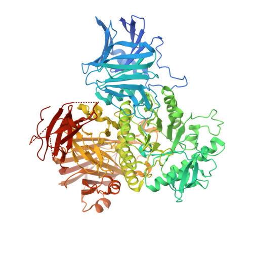
    7F7Q, 7F7R

  • PubMed Abstract: 

    Glycoside hydrolase family 31 (GH31) is a diversified family of anomer-retaining α-glycoside hydrolases, such as α-glucosidase and α-xylosidase, among others. Recently, GH31 α-N-acetylgalactosaminidases (Nag31s) have been identified to hydrolyze the core of mucin-type O-glycans and the crystal structure of a gut bacterium Enterococcus faecalis Nag31 has been reported. However, the mechanisms of substrate specificity and hydrolysis of Nag31s are not well investigated. Herein, we show that E. faecalis Nag31 has the ability to release N-acetylgalactosamine (GalNAc) from O-glycoproteins, such as fetuin and mucin, but has low activity against Tn antigen. Mutational analysis and crystal structures of the Michaelis complexes reveal that residues of the active site work in concert with their conformational changes to act on only α-N-acetylgalactosaminides. Docking simulations using GalNAc-attached peptides suggest that the enzyme mainly recognizes GalNAc and side chains of Ser/Thr, but not strictly other peptide residues. Moreover, quantum mechanics calculations indicate that the enzyme preferred p-nitrophenyl α-N-acetylgalactosaminide to Tn antigen and that the hydrolysis progresses through a conformational itinerary, 4 C 1 1 S 3 4 C 1 , in GalNAc of substrates. Our results provide novel insights into the diversification of the sugar recognition and hydrolytic mechanisms of GH31 enzymes.


  • Organizational Affiliation

    Research Institute of Green Science and Technology, Shizuoka University, 836 Ohya, Suruga-ku, Shizuoka, 422-8529, Japan; Department of Bioscience, Graduate School of Science and Technology, Shizuoka University, 836 Ohya, Suruga-ku, Shizuoka, 422-8529, Japan. Electronic address: miyazaki.takatsugu@shizuoka.ac.jp.


Macromolecules
Find similar proteins by:  (by identity cutoff)  |  3D Structure
Entity ID: 1
MoleculeChains Sequence LengthOrganismDetailsImage
GH31 alpha-N-acetylgalactosaminidase963Enterococcus faecalis ATCC 10100Mutation(s): 0 
Gene Names: WOW_01747
UniProt
Find proteins for Q833V2 (Enterococcus faecalis (strain ATCC 700802 / V583))
Explore Q833V2 
Go to UniProtKB:  Q833V2
Entity Groups  
Sequence Clusters30% Identity50% Identity70% Identity90% Identity95% Identity100% Identity
UniProt GroupQ833V2
Sequence Annotations
Expand
  • Reference Sequence
Small Molecules
Ligands 4 Unique
IDChains Name / Formula / InChI Key2D Diagram3D Interactions
1PE
Query on 1PE

Download Ideal Coordinates CCD File 
K [auth A]PENTAETHYLENE GLYCOL
C10 H22 O6
JLFNLZLINWHATN-UHFFFAOYSA-N
A2G (Subject of Investigation/LOI)
Query on A2G

Download Ideal Coordinates CCD File 
B [auth A]2-acetamido-2-deoxy-alpha-D-galactopyranose
C8 H15 N O6
OVRNDRQMDRJTHS-CBQIKETKSA-N
NPO (Subject of Investigation/LOI)
Query on NPO

Download Ideal Coordinates CCD File 
C [auth A]P-NITROPHENOL
C6 H5 N O3
BTJIUGUIPKRLHP-UHFFFAOYSA-N
EDO
Query on EDO

Download Ideal Coordinates CCD File 
D [auth A]
E [auth A]
F [auth A]
G [auth A]
H [auth A]
D [auth A],
E [auth A],
F [auth A],
G [auth A],
H [auth A],
I [auth A],
J [auth A],
L [auth A]
1,2-ETHANEDIOL
C2 H6 O2
LYCAIKOWRPUZTN-UHFFFAOYSA-N
Experimental Data & Validation

Experimental Data

  • Method: X-RAY DIFFRACTION
  • Resolution: 1.42 Å
  • R-Value Free: 0.196 
  • R-Value Work: 0.174 
  • Space Group: P 21 21 21
Unit Cell:
Length ( Å )Angle ( ˚ )
a = 81.875α = 90
b = 83.394β = 90
c = 146.329γ = 90
Software Package:
Software NamePurpose
REFMACrefinement
XDSdata reduction
SCALAdata scaling
MOLREPphasing
Cootmodel building

Structure Validation

View Full Validation Report


Ligand Structure Quality Assessment 


Entry History & Funding Information

Deposition Data

  • Released Date: 2021-12-08 
  • Deposition Author(s): Miyazaki, T.

Funding OrganizationLocationGrant Number
Japan Society for the Promotion of Science (JSPS)Japan19K15748

Revision History  (Full details and data files)

  • Version 1.0: 2021-12-08
    Type: Initial release
  • Version 1.1: 2022-03-30
    Changes: Database references
  • Version 1.2: 2023-11-29
    Changes: Data collection, Derived calculations, Refinement description