


Molecular basis of dimerization of lytic transglycosylase revealed by the crystal structure of MltA from Acinetobacter baumannii
Jang, H.S., Do, H., Kim, C.M., Kim, G.E., Lee, J.H., Park, H.H.(2021) IUCrJ 8: 921-930
Experimental Data Snapshot
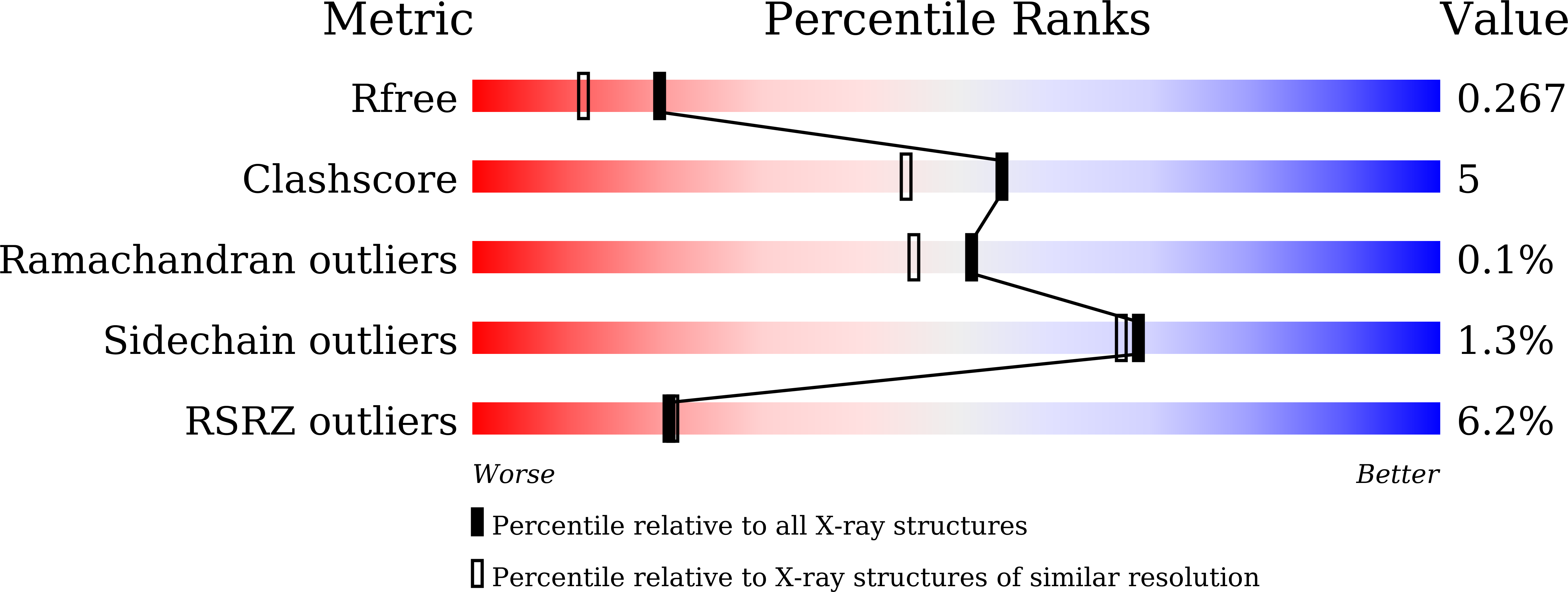
wwPDB Validation 3D Report Full Report
(2021) IUCrJ 8: 921-930
Entity ID: 1 | |||||
|---|---|---|---|---|---|
| Molecule | Chains | Sequence Length | Organism | Details | Image |
| membrane-bound lytic murein transglycosylase A | 366 | Acinetobacter baumannii | Mutation(s): 0 | 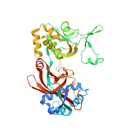 | |
Entity Groups | |||||
| Sequence Clusters | 30% Identity50% Identity70% Identity90% Identity95% Identity100% Identity | ||||
Sequence AnnotationsExpand | |||||
| |||||
| Length ( Å ) | Angle ( ˚ ) |
|---|---|
| a = 97.73 | α = 90 |
| b = 67.44 | β = 98.87 |
| c = 142.37 | γ = 90 |
| Software Name | Purpose |
|---|---|
| PHENIX | refinement |
| PDB_EXTRACT | data extraction |
| HKL-2000 | data collection |
| XDS | data scaling |
| Arcimboldo | phasing |
| Funding Organization | Location | Grant Number |
|---|---|---|
| National Research Foundation (NRF, Korea) | Korea, Republic Of | -- |
RCSB PDB (citation) is hosted by
RCSB PDB is a member of the

