7DF6

Mouse Galectin-3 CRD in complex with novel tetrahydropyran-based thiodisaccharide mimic inhibitor


Experimental Data Snapshot

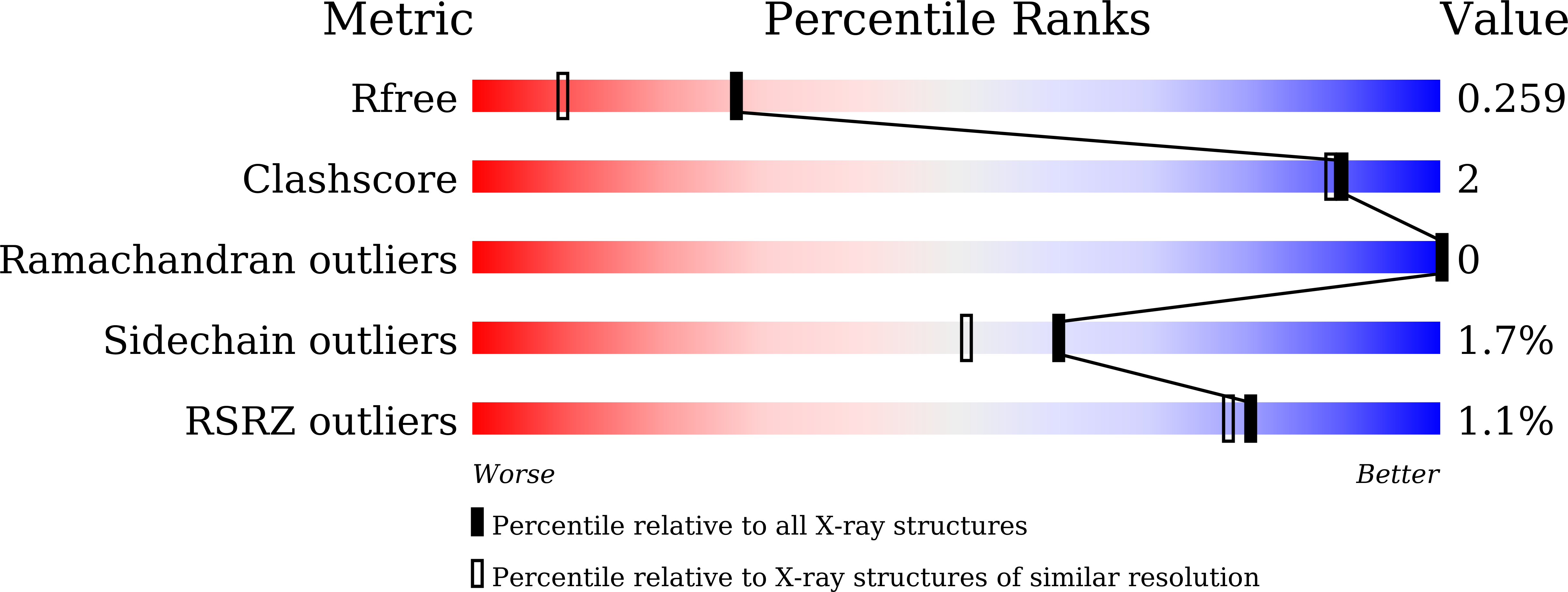
  • Method: X-RAY DIFFRACTION
  • Resolution: 1.80 Å
  • R-Value Free: 0.255 
  • R-Value Work: 0.190 
  • R-Value Observed: 0.193 

wwPDB Validation   3D Report Full Report


Ligand Structure Quality Assessment 


This is version 1.1 of the entry. See complete history


Literature

Synthesis, Structure-Activity Relationships, and In Vivo Evaluation of Novel Tetrahydropyran-Based Thiodisaccharide Mimics as Galectin-3 Inhibitors.

Xu, L.Hartz, R.A.Beno, B.R.Ghosh, K.Shukla, J.K.Kumar, A.Patel, D.Kalidindi, N.Lemos, N.Gautam, S.S.Kumar, A.Ellsworth, B.A.Shah, D.Sale, H.Cheng, D.Regueiro-Ren, A.

(2021) J Med Chem 64: 6634-6655

  • DOI: https://doi.org/10.1021/acs.jmedchem.0c02001
  • Primary Citation of Related Structures:  
    7DF5, 7DF6

  • PubMed Abstract: 

    Galectin-3 is a member of a family of β-galactoside-binding proteins. A substantial body of literature reports that galectin-3 plays important roles in cancer, inflammation, and fibrosis. Small-molecule galectin-3 inhibitors, which are generally lactose or galactose-based derivatives, have the potential to be valuable disease-modifying agents. In our efforts to identify novel galectin-3 disaccharide mimics to improve drug-like properties, we found that one of the monosaccharide subunits can be replaced with a suitably functionalized tetrahydropyran ring. Optimization of the structure-activity relationships around the tetrahydropyran-based scaffold led to the discovery of potent galectin-3 inhibitors. Compounds 36 , 40 , and 45 were selected for further in vivo evaluation. The synthesis, structure-activity relationships, and in vivo evaluation of novel tetrahydropyran-based galectin-3 inhibitors are described.


  • Organizational Affiliation

    Department of Small Molecule Drug Discovery, Bristol Myers Squibb Company, Research and Development, P.O. Box 5400, Princeton, New Jersey 08543, United States.


Macromolecules
Find similar proteins by:  (by identity cutoff)  |  3D Structure
Entity ID: 1
MoleculeChains Sequence LengthOrganismDetailsImage
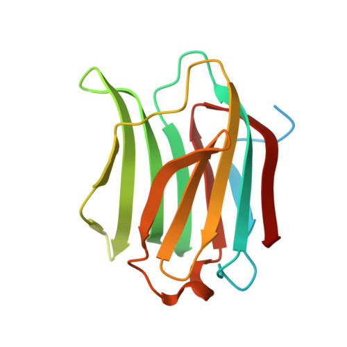
Galectin-3
A, B
165Mus musculusMutation(s): 0 
Gene Names: Lgals3
UniProt
Find proteins for P16110 (Mus musculus)
Explore P16110 
Go to UniProtKB:  P16110
Entity Groups  
Sequence Clusters30% Identity50% Identity70% Identity90% Identity95% Identity100% Identity
UniProt GroupP16110
Sequence Annotations
Expand
  • Reference Sequence
Small Molecules
Ligands 1 Unique
IDChains Name / Formula / InChI Key2D Diagram3D Interactions
H5O (Subject of Investigation/LOI)
Query on H5O

Download Ideal Coordinates CCD File 
C [auth A],
D [auth B]
(2R,3R,4S,5R,6S)-2-(hydroxymethyl)-5-methoxy-6-[(3R,4R,5S)-4-oxidanyl-5-(4-pyrimidin-5-yl-1,2,3-triazol-1-yl)oxan-3-yl]sulfanyl-4-[4-[3,4,5-tris(fluoranyl)phenyl]-1,2,3-triazol-1-yl]oxan-3-ol
C26 H27 F3 N8 O6 S
XUWUKOCAMCFANQ-COQUQQDQSA-N
Experimental Data & Validation

Experimental Data

  • Method: X-RAY DIFFRACTION
  • Resolution: 1.80 Å
  • R-Value Free: 0.255 
  • R-Value Work: 0.190 
  • R-Value Observed: 0.193 
  • Space Group: P 21 21 21
Unit Cell:
Length ( Å )Angle ( ˚ )
a = 36.14α = 90
b = 58.14β = 90
c = 130.06γ = 90
Software Package:
Software NamePurpose
REFMACrefinement
Aimlessdata scaling
PDB_EXTRACTdata extraction
XDSdata reduction
PHASERphasing

Structure Validation

View Full Validation Report


Ligand Structure Quality Assessment 


Entry History 

Deposition Data

Revision History  (Full details and data files)

  • Version 1.0: 2021-06-23
    Type: Initial release
  • Version 1.1: 2023-11-29
    Changes: Data collection, Database references, Refinement description