7A3R

Crystal structure of dengue 1 virus envelope glycoprotein


Experimental Data Snapshot

  • Method: X-RAY DIFFRACTION
  • Resolution: 3.60 Å
  • R-Value Free: 0.295 
  • R-Value Work: 0.250 
  • R-Value Observed: 0.253 

wwPDB Validation   3D Report Full Report


This is version 1.3 of the entry. See complete history


Literature

The epitope arrangement on flavivirus particles contributes to Mab C10's extraordinary neutralization breadth across Zika and dengue viruses.

Sharma, A.Zhang, X.Dejnirattisai, W.Dai, X.Gong, D.Wongwiwat, W.Duquerroy, S.Rouvinski, A.Vaney, M.C.Guardado-Calvo, P.Haouz, A.England, P.Sun, R.Zhou, Z.H.Mongkolsapaya, J.Screaton, G.R.Rey, F.A.

(2021) Cell 184: 6052-6066.e18

  • DOI: https://doi.org/10.1016/j.cell.2021.11.010
  • Primary Citation of Related Structures:  
    7A3N, 7A3O, 7A3P, 7A3Q, 7A3R, 7A3S, 7A3T, 7A3U, 7CTH

  • PubMed Abstract: 

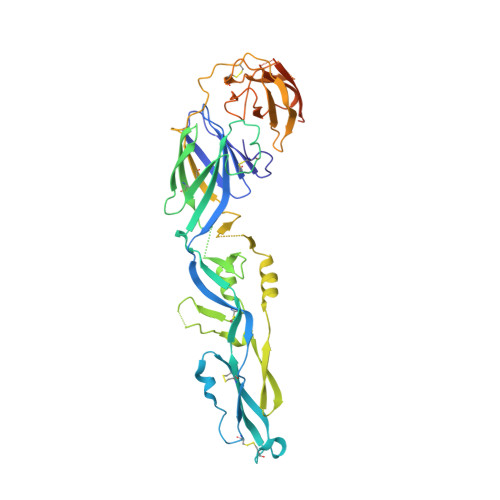
    The human monoclonal antibody C10 exhibits extraordinary cross-reactivity, potently neutralizing Zika virus (ZIKV) and the four serotypes of dengue virus (DENV1-DENV4). Here we describe a comparative structure-function analysis of C10 bound to the envelope (E) protein dimers of the five viruses it neutralizes. We demonstrate that the C10 Fab has high affinity for ZIKV and DENV1 but not for DENV2, DENV3, and DENV4. We further show that the C10 interaction with the latter viruses requires an E protein conformational landscape that limits binding to only one of the three independent epitopes per virion. This limited affinity is nevertheless counterbalanced by the particle's icosahedral organization, which allows two different dimers to be reached by both Fab arms of a C10 immunoglobulin. The epitopes' geometric distribution thus confers C10 its exceptional neutralization breadth. Our results highlight the importance not only of paratope/epitope complementarity but also the topological distribution for epitope-focused vaccine design.


  • Organizational Affiliation

    Institut Pasteur, Université de Paris, CNRS UMR3569, Unité de Virologie Structurale, 75015 Paris, France.


Macromolecules
Find similar proteins by:  (by identity cutoff)  |  3D Structure
Entity ID: 1
MoleculeChains Sequence LengthOrganismDetailsImage
Core protein
A, B
433dengue virus type 1Mutation(s): 0 
EC: 3.4.21.91 (PDB Primary Data), 3.6.1.15 (PDB Primary Data), 3.6.4.13 (PDB Primary Data)
UniProt
Find proteins for B1N6C5 (dengue virus type 1)
Explore B1N6C5 
Go to UniProtKB:  B1N6C5
Entity Groups  
Sequence Clusters30% Identity50% Identity70% Identity90% Identity95% Identity100% Identity
UniProt GroupB1N6C5
Sequence Annotations
Expand
  • Reference Sequence
Oligosaccharides

Help

Entity ID: 2
MoleculeChains Length2D Diagram Glycosylation3D Interactions
alpha-L-fucopyranose-(1-6)-2-acetamido-2-deoxy-beta-D-glucopyranose
C
2N-Glycosylation
Glycosylation Resources
GlyTouCan:  G86851RC
GlyCosmos:  G86851RC
GlyGen:  G86851RC
Small Molecules
Ligands 1 Unique
IDChains Name / Formula / InChI Key2D Diagram3D Interactions
NAG
Query on NAG

Download Ideal Coordinates CCD File 
D [auth A],
E [auth B]
2-acetamido-2-deoxy-beta-D-glucopyranose
C8 H15 N O6
OVRNDRQMDRJTHS-FMDGEEDCSA-N
Experimental Data & Validation

Experimental Data

  • Method: X-RAY DIFFRACTION
  • Resolution: 3.60 Å
  • R-Value Free: 0.295 
  • R-Value Work: 0.250 
  • R-Value Observed: 0.253 
  • Space Group: P 41 21 2
Unit Cell:
Length ( Å )Angle ( ˚ )
a = 105.19α = 90
b = 105.19β = 90
c = 180.04γ = 90
Software Package:
Software NamePurpose
PHENIXrefinement
PDB_EXTRACTdata extraction
Aimlessdata scaling
PHASERphasing
XDSdata reduction

Structure Validation

View Full Validation Report


Entry History & Funding Information

Deposition Data


Funding OrganizationLocationGrant Number
Wellcome TrustFrance--

Revision History  (Full details and data files)

  • Version 1.0: 2021-12-08
    Type: Initial release
  • Version 1.1: 2021-12-15
    Changes: Database references
  • Version 1.2: 2021-12-22
    Changes: Database references
  • Version 1.3: 2024-01-31
    Changes: Data collection, Refinement description