6Z2K

The structure of the tetrameric HDAC1/MIDEAS/DNTTIP1 MiDAC deacetylase complex


Experimental Data Snapshot

  • Method: ELECTRON MICROSCOPY
  • Resolution: 4.50 Å
  • Aggregation State: PARTICLE 
  • Reconstruction Method: SINGLE PARTICLE 

wwPDB Validation   3D Report Full Report


This is version 2.0 of the entry. See complete history


Literature

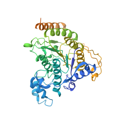
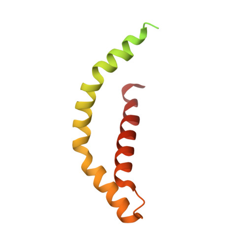
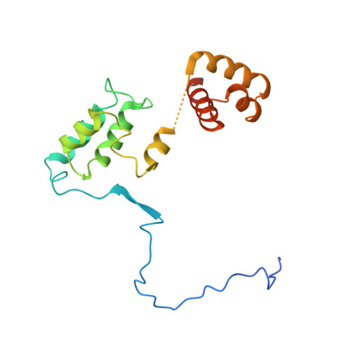
The MiDAC histone deacetylase complex is essential for embryonic development and has a unique multivalent structure.

Turnbull, R.E.Fairall, L.Saleh, A.Kelsall, E.Morris, K.L.Ragan, T.J.Savva, C.G.Chandru, A.Millard, C.J.Makarova, O.V.Smith, C.J.Roseman, A.M.Fry, A.M.Cowley, S.M.Schwabe, J.W.R.

(2020) Nat Commun 11: 3252-3252

  • DOI: https://doi.org/10.1038/s41467-020-17078-8
  • Primary Citation of Related Structures:  
    6Z2J, 6Z2K

  • PubMed Abstract: 

    MiDAC is one of seven distinct, large multi-protein complexes that recruit class I histone deacetylases to the genome to regulate gene expression. Despite implications of involvement in cell cycle regulation and in several cancers, surprisingly little is known about the function or structure of MiDAC. Here we show that MiDAC is important for chromosome alignment during mitosis in cancer cell lines. Mice lacking the MiDAC proteins, DNTTIP1 or MIDEAS, die with identical phenotypes during late embryogenesis due to perturbations in gene expression that result in heart malformation and haematopoietic failure. This suggests that MiDAC has an essential and unique function that cannot be compensated by other HDAC complexes. Consistent with this, the cryoEM structure of MiDAC reveals a unique and distinctive mode of assembly. Four copies of HDAC1 are positioned at the periphery with outward-facing active sites suggesting that the complex may target multiple nucleosomes implying a processive deacetylase function.


  • Organizational Affiliation

    Leicester Institute of Structural and Chemical Biology, University of Leicester, Leicester, LE1 7RH, UK.


Macromolecules
Find similar proteins by:  (by identity cutoff)  |  3D Structure
Entity ID: 1
MoleculeChains Sequence LengthOrganismDetailsImage
Histone deacetylase 1A [auth C],
B [auth E],
C [auth K],
D [auth I]
482Homo sapiensMutation(s): 0 
Gene Names: HDAC1RPD3L1
EC: 3.5.1.98
UniProt & NIH Common Fund Data Resources
Find proteins for Q13547 (Homo sapiens)
Explore Q13547 
Go to UniProtKB:  Q13547
PHAROS:  Q13547
GTEx:  ENSG00000116478 
Entity Groups  
Sequence Clusters30% Identity50% Identity70% Identity90% Identity95% Identity100% Identity
UniProt GroupQ13547
Sequence Annotations
Expand
  • Reference Sequence
Find similar proteins by:  (by identity cutoff)  |  3D Structure
Entity ID: 2
MoleculeChains Sequence LengthOrganismDetailsImage
Deoxynucleotidyltransferase terminal-interacting protein 1E [auth A],
F [auth B],
G,
H
130Homo sapiensMutation(s): 0 
Gene Names: DNTTIP1C20orf167TDIF1
UniProt & NIH Common Fund Data Resources
Find proteins for Q9H147 (Homo sapiens)
Explore Q9H147 
Go to UniProtKB:  Q9H147
PHAROS:  Q9H147
GTEx:  ENSG00000101457 
Entity Groups  
Sequence Clusters30% Identity50% Identity70% Identity90% Identity95% Identity100% Identity
UniProt GroupQ9H147
Sequence Annotations
Expand
  • Reference Sequence
Find similar proteins by:  (by identity cutoff)  |  3D Structure
Entity ID: 3
MoleculeChains Sequence LengthOrganismDetailsImage
Mitotic deacetylase-associated SANT domain proteinI [auth F],
J [auth D],
K [auth L],
L [auth J]
173Homo sapiensMutation(s): 0 
Gene Names: MIDEASC14orf117C14orf43ELMSAN1
UniProt & NIH Common Fund Data Resources
Find proteins for Q6PJG2 (Homo sapiens)
Explore Q6PJG2 
Go to UniProtKB:  Q6PJG2
PHAROS:  Q6PJG2
GTEx:  ENSG00000156030 
Entity Groups  
Sequence Clusters30% Identity50% Identity70% Identity90% Identity95% Identity100% Identity
UniProt GroupQ6PJG2
Sequence Annotations
Expand
  • Reference Sequence
Small Molecules
Ligands 3 Unique
IDChains Name / Formula / InChI Key2D Diagram3D Interactions
IHP
Query on IHP

Download Ideal Coordinates CCD File 
AA [auth D],
BA [auth J],
P [auth E],
Z [auth F]
INOSITOL HEXAKISPHOSPHATE
C6 H18 O24 P6
IMQLKJBTEOYOSI-GPIVLXJGSA-N
ZN
Query on ZN

Download Ideal Coordinates CCD File 
M [auth C],
Q [auth E],
T [auth K],
W [auth I]
ZINC ION
Zn
PTFCDOFLOPIGGS-UHFFFAOYSA-N
K
Query on K

Download Ideal Coordinates CCD File 
N [auth C]
O [auth C]
R [auth E]
S [auth E]
U [auth K]
N [auth C],
O [auth C],
R [auth E],
S [auth E],
U [auth K],
V [auth K],
X [auth I],
Y [auth I]
POTASSIUM ION
K
NPYPAHLBTDXSSS-UHFFFAOYSA-N
Experimental Data & Validation

Experimental Data

  • Method: ELECTRON MICROSCOPY
  • Resolution: 4.50 Å
  • Aggregation State: PARTICLE 
  • Reconstruction Method: SINGLE PARTICLE 
EM Software:
TaskSoftware PackageVersion
MODEL REFINEMENTPHENIX1.14
RECONSTRUCTIONRELION3.0

Structure Validation

View Full Validation Report


Entry History & Funding Information

Deposition Data


Funding OrganizationLocationGrant Number
Wellcome TrustUnited Kingdom100237/Z/12/Z
Biotechnology and Biological Sciences Research Council (BBSRC)United KingdomBB/N002954/1
Medical Research Council (MRC, United Kingdom)United KingdomMC_PC_171136

Revision History  (Full details and data files)

  • Version 1.0: 2020-07-08
    Type: Initial release
  • Version 2.0: 2020-10-07
    Changes: Advisory, Atomic model, Data collection, Derived calculations, Non-polymer description, Structure summary