


Crystal Structure of Serine protease SplB N2K/N3Q/S154R from Staphylococcus aureus
Knyphausen, P., Rangel Pereira, M.R., Brear, P., Jermutus, L., Hollfelder, F.To be published.
Experimental Data Snapshot
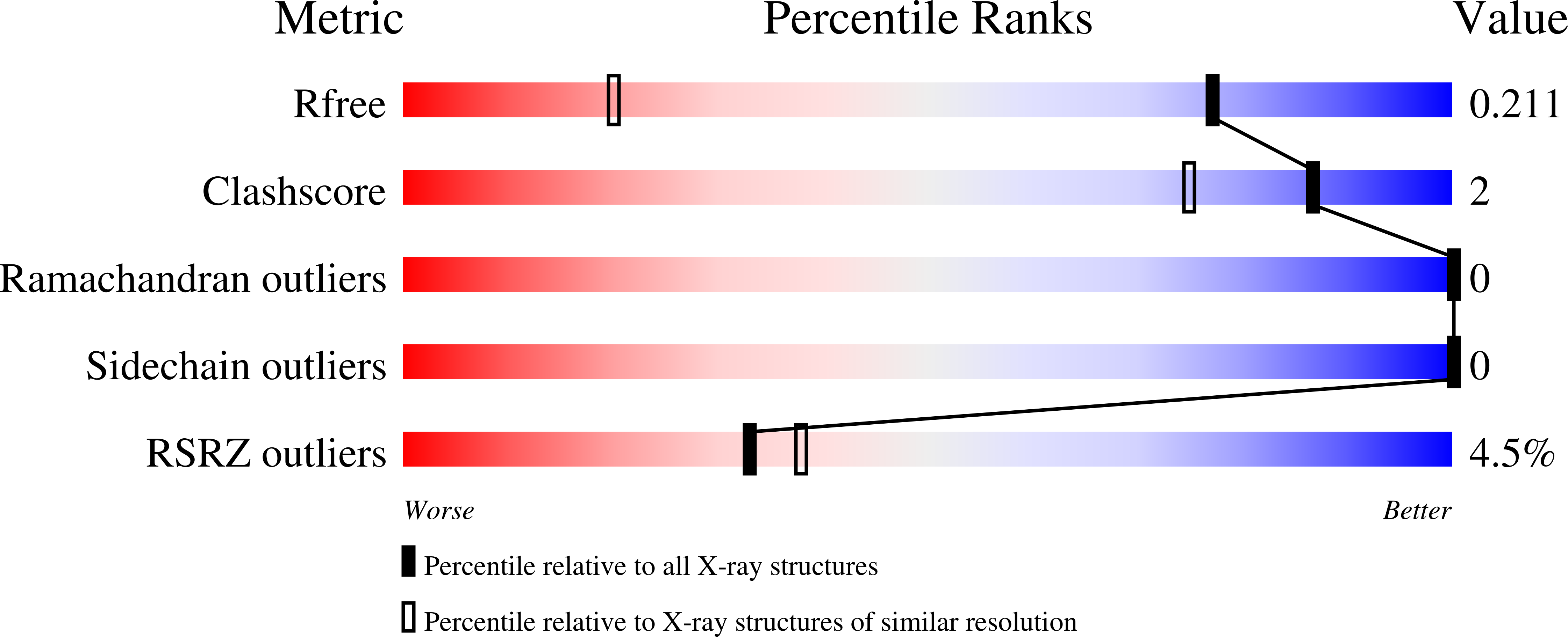
wwPDB Validation 3D Report Full Report
Entity ID: 1 | |||||
|---|---|---|---|---|---|
| Molecule | Chains | Sequence Length | Organism | Details | Image |
| Serine protease | 206 | Staphylococcus aureus | Mutation(s): 3 Gene Names: splB EC: 3.4.21 | 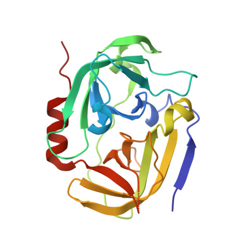 | |
UniProt | |||||
Find proteins for Q2FXC3 (Staphylococcus aureus (strain NCTC 8325 / PS 47)) Explore Q2FXC3 Go to UniProtKB: Q2FXC3 | |||||
Entity Groups | |||||
| Sequence Clusters | 30% Identity50% Identity70% Identity90% Identity95% Identity100% Identity | ||||
| UniProt Group | Q2FXC3 | ||||
Sequence AnnotationsExpand | |||||
| |||||
| Ligands 2 Unique | |||||
|---|---|---|---|---|---|
| ID | Chains | Name / Formula / InChI Key | 2D Diagram | 3D Interactions | |
| 1PE Query on 1PE | B [auth A], C [auth A] | PENTAETHYLENE GLYCOL C10 H22 O6 JLFNLZLINWHATN-UHFFFAOYSA-N |  | ||
| PEG Query on PEG | D [auth A] | DI(HYDROXYETHYL)ETHER C4 H10 O3 MTHSVFCYNBDYFN-UHFFFAOYSA-N |  | ||
| Length ( Å ) | Angle ( ˚ ) |
|---|---|
| a = 130.69 | α = 90 |
| b = 44.31 | β = 92.94 |
| c = 30.81 | γ = 90 |
| Software Name | Purpose |
|---|---|
| XDS | data reduction |
| Aimless | data scaling |
| PHASER | phasing |
| BUSTER | refinement |
| PDB_EXTRACT | data extraction |
| Funding Organization | Location | Grant Number |
|---|---|---|
| European Research Council (ERC) | United Kingdom | 695669 |
RCSB PDB (citation) is hosted by
RCSB PDB is a member of the

