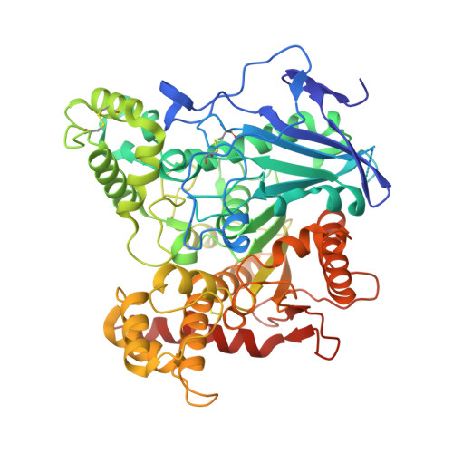
A Second Look at the Crystal Structures ofDrosophila melanogasterAcetylcholinesterase in Complex with Tacrine Derivatives Provides Insights Concerning Catalytic Intermediates and the Design of Specific Insecticides.
Nachon, F., Rosenberry, T.L., Silman, I., Sussman, J.L.(2020) Molecules 25
- PubMed: 32155891
- DOI: https://doi.org/10.3390/molecules25051198
- Primary Citation of Related Structures:
6XYS, 6XYU, 6XYY - PubMed Abstract:
Over recent decades, crystallographic software for data processing and structure refinement has improved dramatically, resulting in more accurate and detailed crystal structures. It is, therefore, sometimes valuable to have a second look at "old" diffraction data, especially when earlier interpretation of the electron density maps was rather difficult. Here, we present updated crystal structures of Drosophila melanogaster acetylcholinesterase ( Dm AChE) originally published in [Harel et al., Prot Sci (2000) 9:1063-1072], which reveal features previously unnoticed. Thus, previously unmodeled density in the native active site can be interpreted as stable acetylation of the catalytic serine. Similarly, a strong density in the Dm AChE/ZA complex originally attributed to a sulfate ion is better interpreted as a small molecule that is covalently bound. This small molecule can be modeled as either a propionate or a glycinate. The complex is reminiscent of the carboxylate butyrylcholinesterase complexes observed in crystal structures of human butyrylcholinesterases from various sources, and demonstrates the remarkable ability of cholinesterases to stabilize covalent complexes with carboxylates. A very strong peak of density (10 σ) at covalent distance from the Cβ of the catalytic serine is present in the Dm AChE/ZAI complex. This can be undoubtedly attributed to an iodine atom, suggesting an unanticipated iodo/hydroxyl exchange between Ser238 and the inhibitor, possibly driven by the intense X-ray irradiation. Finally, the binding of tacrine-derived inhibitors, such as ZA (1DX4) or the iodinated analog, ZAI (1QON) results in the appearance of an open channel that connects the base of the active-site gorge to the solvent. This channel, which arises due to the absence of the conserved tyrosine present in vertebrate cholinesterases, could be exploited to design inhibitors specific to insect cholinesterases. The present study demonstrates that updated processing of older diffraction images, and the re-refinement of older diffraction data, can produce valuable information that could not be detected in the original analysis, and strongly supports the preservation of the diffraction images in public data banks.
Organizational Affiliation:
Département de Toxicologie et Risques Chimiques, Institut de Recherche Biomédicale des Armées, 91220 Brétigny-sur-Orge, France.