6XB9

Crystal structure of Azotobacter vinelandii 3-mercaptopropionic acid dioxygenase in complex with 3-hydroxypropionic acid


Experimental Data Snapshot

  • Method: X-RAY DIFFRACTION
  • Resolution: 2.25 Å
  • R-Value Free: 0.226 
  • R-Value Work: 0.196 
  • R-Value Observed: 0.197 

wwPDB Validation   3D Report Full Report


Ligand Structure Quality Assessment 


This is version 1.3 of the entry. See complete history


Literature

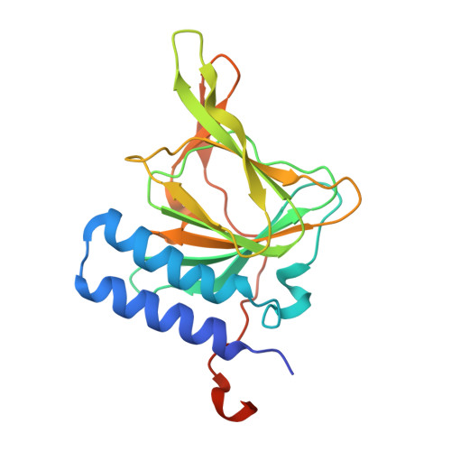
Structure of 3-mercaptopropionic acid dioxygenase with a substrate analog reveals bidentate substrate binding at the iron center.

York, N.J.Lockart, M.M.Sardar, S.Khadka, N.Shi, W.Stenkamp, R.E.Zhang, J.Kiser, P.D.Pierce, B.S.

(2021) J Biol Chem 296: 100492-100492

  • DOI: https://doi.org/10.1016/j.jbc.2021.100492
  • Primary Citation of Related Structures:  
    6XB9, 7KOV

  • PubMed Abstract: 

    Thiol dioxygenases are a subset of nonheme iron oxygenases that catalyze the formation of sulfinic acids from sulfhydryl-containing substrates and dioxygen. Among this class, cysteine dioxygenases (CDOs) and 3-mercaptopropionic acid dioxygenases (3MDOs) are the best characterized, and the mode of substrate binding for CDOs is well understood. However, the manner in which 3-mercaptopropionic acid (3MPA) coordinates to the nonheme iron site in 3MDO remains a matter of debate. A model for bidentate 3MPA coordination at the 3MDO Fe-site has been proposed on the basis of computational docking, whereas steady-state kinetics and EPR spectroscopic measurements suggest a thiolate-only coordination of the substrate. To address this gap in knowledge, we determined the structure of Azobacter vinelandii 3MDO (Av3MDO) in complex with the substrate analog and competitive inhibitor, 3-hydroxypropionic acid (3HPA). The structure together with DFT computational modeling demonstrates that 3HPA and 3MPA associate with iron as chelate complexes with the substrate-carboxylate group forming an additional interaction with Arg168 and the thiol bound at the same position as in CDO. A chloride ligand was bound to iron in the coordination site assigned as the O 2 -binding site. Supporting HYSCORE spectroscopic experiments were performed on the (3MPA/NO)-bound Av3MDO iron nitrosyl (S = 3/2) site. In combination with spectroscopic simulations and optimized DFT models, this work provides an experimentally verified model of the Av3MDO enzyme-substrate complex, effectively resolving a debate in the literature regarding the preferred substrate-binding denticity. These results elegantly explain the observed 3MDO substrate specificity, but leave unanswered questions regarding the mechanism of substrate-gated reactivity with dioxygen.


  • Organizational Affiliation

    Department of Chemistry & Biochemistry, University of Alabama, Tuscaloosa, Alabama, USA.


Macromolecules
Find similar proteins by:  (by identity cutoff)  |  3D Structure
Entity ID: 1
MoleculeChains Sequence LengthOrganismDetailsImage
Cysteine dioxygenase type I protein
A, B, C, D, E
A, B, C, D, E, F, G, H, I, J, K, L
208Azotobacter vinelandiiMutation(s): 0 
UniProt
Find proteins for C1DN94 (Azotobacter vinelandii (strain DJ / ATCC BAA-1303))
Explore C1DN94 
Go to UniProtKB:  C1DN94
Entity Groups  
Sequence Clusters30% Identity50% Identity70% Identity90% Identity95% Identity100% Identity
UniProt GroupC1DN94
Sequence Annotations
Expand
  • Reference Sequence
Small Molecules
Ligands 4 Unique
IDChains Name / Formula / InChI Key2D Diagram3D Interactions
3OH (Subject of Investigation/LOI)
Query on 3OH

Download Ideal Coordinates CCD File 
BA [auth E]
EA [auth F]
HA [auth G]
KA [auth H]
N [auth A]
BA [auth E],
EA [auth F],
HA [auth G],
KA [auth H],
N [auth A],
PA [auth I],
R [auth B],
SA [auth J],
U [auth C],
WA [auth K],
Y [auth D],
ZA [auth L]
3-HYDROXY-PROPANOIC ACID
C3 H6 O3
ALRHLSYJTWAHJZ-UHFFFAOYSA-N
FE (Subject of Investigation/LOI)
Query on FE

Download Ideal Coordinates CCD File 
AA [auth E]
DA [auth F]
GA [auth G]
JA [auth H]
M [auth A]
AA [auth E],
DA [auth F],
GA [auth G],
JA [auth H],
M [auth A],
OA [auth I],
Q [auth B],
RA [auth J],
T [auth C],
VA [auth K],
X [auth D],
YA [auth L]
FE (III) ION
Fe
VTLYFUHAOXGGBS-UHFFFAOYSA-N
CL (Subject of Investigation/LOI)
Query on CL

Download Ideal Coordinates CCD File 
AB [auth L]
CA [auth E]
FA [auth F]
IA [auth G]
LA [auth H]
AB [auth L],
CA [auth E],
FA [auth F],
IA [auth G],
LA [auth H],
MA [auth H],
O [auth A],
QA [auth I],
S [auth B],
TA [auth J],
V [auth C],
XA [auth K],
Z [auth D]
CHLORIDE ION
Cl
VEXZGXHMUGYJMC-UHFFFAOYSA-M
MG
Query on MG

Download Ideal Coordinates CCD File 
NA [auth H],
P [auth A],
UA [auth J],
W [auth C]
MAGNESIUM ION
Mg
JLVVSXFLKOJNIY-UHFFFAOYSA-N
Experimental Data & Validation

Experimental Data

  • Method: X-RAY DIFFRACTION
  • Resolution: 2.25 Å
  • R-Value Free: 0.226 
  • R-Value Work: 0.196 
  • R-Value Observed: 0.197 
  • Space Group: P 31
Unit Cell:
Length ( Å )Angle ( ˚ )
a = 178.219α = 90
b = 178.219β = 90
c = 75.915γ = 120
Software Package:
Software NamePurpose
REFMACrefinement
PDB_EXTRACTdata extraction
XDSdata reduction
XDSdata scaling
PHASERphasing

Structure Validation

View Full Validation Report


Ligand Structure Quality Assessment 


Entry History 

Deposition Data

Revision History  (Full details and data files)

  • Version 1.0: 2021-03-03
    Type: Initial release
  • Version 1.1: 2021-03-17
    Changes: Database references
  • Version 1.2: 2021-07-14
    Changes: Database references
  • Version 1.3: 2023-10-18
    Changes: Data collection, Database references, Refinement description