6W70

Crystal Structure of apixaban-bound ABLE


Experimental Data Snapshot

  • Method: X-RAY DIFFRACTION
  • Resolution: 1.30 Å
  • R-Value Free: 0.183 
  • R-Value Work: 0.161 
  • R-Value Observed: 0.162 

wwPDB Validation   3D Report Full Report


Ligand Structure Quality Assessment 


This is version 1.3 of the entry. See complete history


Literature

A defined structural unit enables de novo design of small-molecule-binding proteins.

Polizzi, N.F.DeGrado, W.F.

(2020) Science 369: 1227-1233

  • DOI: https://doi.org/10.1126/science.abb8330
  • Primary Citation of Related Structures:  
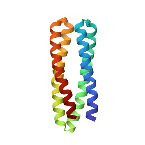
    6W6X, 6W70, 6X8N

  • PubMed Abstract: 

    The de novo design of proteins that bind highly functionalized small molecules represents a great challenge. To enable computational design of binders, we developed a unit of protein structure-a van der Mer (vdM)-that maps the backbone of each amino acid to statistically preferred positions of interacting chemical groups. Using vdMs, we designed six de novo proteins to bind the drug apixaban; two bound with low and submicromolar affinity. X-ray crystallography and mutagenesis confirmed a structure with a precisely designed cavity that forms favorable interactions in the drug-protein complex. vdMs may enable design of functional proteins for applications in sensing, medicine, and catalysis.


  • Organizational Affiliation

    Department of Pharmaceutical Chemistry, Cardiovascular Research Institute, University of California, San Francisco, San Francisco, CA 94158, USA. nicholas.polizzi@ucsf.edu william.degrado@ucsf.edu.


Macromolecules
Find similar proteins by:  (by identity cutoff)  |  3D Structure
Entity ID: 1
MoleculeChains Sequence LengthOrganismDetailsImage
De novo designed ABLEA,
B [auth C]
126synthetic constructMutation(s): 0 
Entity Groups  
Sequence Clusters30% Identity50% Identity70% Identity90% Identity95% Identity100% Identity
Sequence Annotations
Expand
  • Reference Sequence
Experimental Data & Validation

Experimental Data

  • Method: X-RAY DIFFRACTION
  • Resolution: 1.30 Å
  • R-Value Free: 0.183 
  • R-Value Work: 0.161 
  • R-Value Observed: 0.162 
  • Space Group: P 1 21 1
Unit Cell:
Length ( Å )Angle ( ˚ )
a = 35.194α = 90
b = 78.445β = 106.95
c = 43.133γ = 90
Software Package:
Software NamePurpose
PHENIXrefinement
SCALAdata scaling
PDB_EXTRACTdata extraction
XDSdata reduction
PHENIXphasing

Structure Validation

View Full Validation Report


Ligand Structure Quality Assessment 


Entry History & Funding Information

Deposition Data


Funding OrganizationLocationGrant Number
National Institutes of Health/National Institute of General Medical Sciences (NIH/NIGMS)--

Revision History  (Full details and data files)

  • Version 1.0: 2020-08-26
    Type: Initial release
  • Version 1.1: 2020-09-23
    Changes: Database references
  • Version 1.2: 2024-03-06
    Changes: Data collection, Database references
  • Version 1.3: 2024-04-03
    Changes: Refinement description