6W1I

Re-interpretation of ppGpp (G4P) electron density in the deposited crystal structure of Xanthine phosphoribosyltransferase (XPRT) (1Y0B).


Experimental Data Snapshot

  • Method: X-RAY DIFFRACTION
  • Resolution: 1.80 Å
  • R-Value Free: 0.208 
  • R-Value Work: 0.160 
  • R-Value Observed: 0.162 

wwPDB Validation   3D Report Full Report


Ligand Structure Quality Assessment 


This is version 1.1 of the entry. See complete history

Re-refinement Note

This entry reflects an alternative modeling of the original data in: 1Y0B


Literature

Molecular Mechanism of Regulation of the Purine Salvage Enzyme XPRT by the Alarmones pppGpp, ppGpp, and pGpp.

Anderson, B.W.Hao, A.Satyshur, K.A.Keck, J.L.Wang, J.D.

(2020) J Mol Biol 432: 4108-4126

  • DOI: https://doi.org/10.1016/j.jmb.2020.05.013
  • Primary Citation of Related Structures:  
    6W1I

  • PubMed Abstract: 

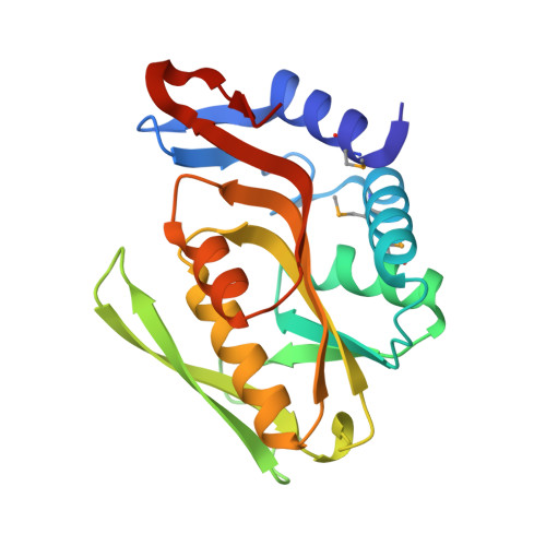
    The alarmones pppGpp and ppGpp mediate starvation response and maintain purine homeostasis to protect bacteria. In the bacterial phyla Firmicutes and Bacteroidetes, xanthine phosphoribosyltransferase (XPRT) is a purine salvage enzyme that produces the nucleotide XMP from PRPP and xanthine. Combining structural, biochemical, and genetic analyses, we show that pppGpp and ppGpp, as well as a third newly identified alarmone pGpp, all directly interact with XPRT from the Gram-positive bacterium Bacillus subtilis and inhibit XPRT activity by competing with its substrate PRPP. Structural analysis reveals that ppGpp binds the PRPP binding motif within the XPRT active site. This motif is present in another (p)ppGpp target, the purine salvage enzyme HPRT, suggesting evolutionary conservation in different enzymes. However, XPRT oligomeric interaction is distinct from HPRT in that XPRT forms a symmetric dimer with two (p)ppGpp binding sites at the dimer interface. (p)ppGpp's interaction with an XPRT bridging loop across the interface results in XPRT cooperatively binding (p)ppGpp. Also, XPRT displays differential regulation by the alarmones as it is potently inhibited by both ppGpp and pGpp, but only modestly by pppGpp. Lastly, we demonstrate that the alarmones are necessary for protecting GTP homeostasis against excess environmental xanthine in B. subtilis, suggesting that regulation of XPRT is key for regulating the purine salvage pathway.


  • Organizational Affiliation

    Department of Bacteriology, University of Wisconsin-Madison, 1550 Linden Dr., Madison, WI 53706, USA.


Macromolecules
Find similar proteins by:  (by identity cutoff)  |  3D Structure
Entity ID: 1
MoleculeChains Sequence LengthOrganismDetailsImage
Xanthine phosphoribosyltransferase
A, B, C, D
197Bacillus subtilis subsp. subtilis str. 168Mutation(s): 0 
Gene Names: xptBSU22070
EC: 2.4.2.22
UniProt
Find proteins for P42085 (Bacillus subtilis (strain 168))
Explore P42085 
Go to UniProtKB:  P42085
Entity Groups  
Sequence Clusters30% Identity50% Identity70% Identity90% Identity95% Identity100% Identity
UniProt GroupP42085
Sequence Annotations
Expand
  • Reference Sequence
Small Molecules
Ligands 2 Unique
IDChains Name / Formula / InChI Key2D Diagram3D Interactions
G4P (Subject of Investigation/LOI)
Query on G4P

Download Ideal Coordinates CCD File 
G [auth A],
J [auth B],
M [auth C],
P [auth D]
GUANOSINE-5',3'-TETRAPHOSPHATE
C10 H17 N5 O17 P4
BUFLLCUFNHESEH-UUOKFMHZSA-N
NA
Query on NA

Download Ideal Coordinates CCD File 
E [auth A]
F [auth A]
H [auth B]
I [auth B]
K [auth C]
E [auth A],
F [auth A],
H [auth B],
I [auth B],
K [auth C],
L [auth C],
N [auth D],
O [auth D]
SODIUM ION
Na
FKNQFGJONOIPTF-UHFFFAOYSA-N
Modified Residues  1 Unique
IDChains TypeFormula2D DiagramParent
MSE
Query on MSE
A, B, C, D
L-PEPTIDE LINKINGC5 H11 N O2 SeMET
Binding Affinity Annotations 
IDSourceBinding Affinity
G4P Binding MOAD:  6W1I Kd: 950 (nM) from 1 assay(s)
Experimental Data & Validation

Experimental Data

  • Method: X-RAY DIFFRACTION
  • Resolution: 1.80 Å
  • R-Value Free: 0.208 
  • R-Value Work: 0.160 
  • R-Value Observed: 0.162 
  • Space Group: P 1 21 1
Unit Cell:
Length ( Å )Angle ( ˚ )
a = 42.503α = 90
b = 115.902β = 94.3
c = 73.985γ = 90
Software Package:
Software NamePurpose
d*TREKdata reduction
HKL-2000data scaling
SOLVEphasing
SHARPphasing
PHENIXrefinement

Structure Validation

View Full Validation Report


Ligand Structure Quality Assessment 


Entry History & Funding Information

Deposition Data


Funding OrganizationLocationGrant Number
National Institutes of Health/National Institute of General Medical Sciences (NIH/NIGMS)United StatesGM62414
National Institutes of Health/National Institute of General Medical Sciences (NIH/NIGMS)United StatesR35 GM127088
National Science Foundation (NSF, United States)United StatesGRFP DGE-1256259

Revision History  (Full details and data files)

  • Version 1.0: 2020-07-29
    Type: Initial release
  • Version 1.1: 2021-04-07
    Changes: Data collection