


Crystal structure of mouse acyl-CoA thioesterase 7 with CoA
Teakel, S.L., Forwood, J.K.To be published.
Experimental Data Snapshot
wwPDB Validation 3D Report Full Report
Currently 6VFY does not have a validation slider image.
Entity ID: 1 | |||||
|---|---|---|---|---|---|
| Molecule | Chains | Sequence Length | Organism | Details | Image |
| Cytosolic acyl coenzyme A thioester hydrolase | A [auth D], B [auth E], C [auth F] | 338 | Mus musculus | Mutation(s): 0 Gene Names: Acot7, Bach EC: 3.1.2.2 | 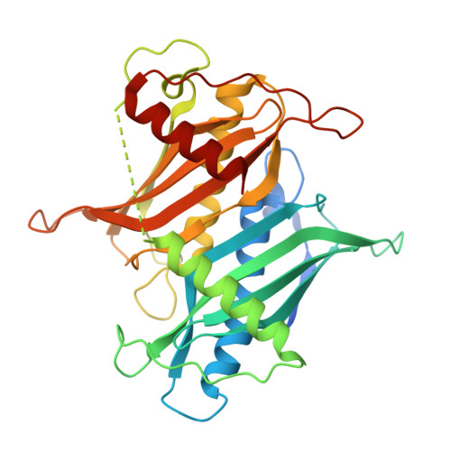 |
UniProt | |||||
Find proteins for Q91V12 (Mus musculus) Explore Q91V12 Go to UniProtKB: Q91V12 | |||||
Entity Groups | |||||
| Sequence Clusters | 30% Identity50% Identity70% Identity90% Identity95% Identity100% Identity | ||||
| UniProt Group | Q91V12 | ||||
Sequence AnnotationsExpand | |||||
| |||||
| Ligands 2 Unique | |||||
|---|---|---|---|---|---|
| ID | Chains | Name / Formula / InChI Key | 2D Diagram | 3D Interactions | |
| COA (Subject of Investigation/LOI) Query on COA | D, E, F | COENZYME A C21 H36 N7 O16 P3 S RGJOEKWQDUBAIZ-IBOSZNHHSA-N |  | ||
| PO4 Query on PO4 | G [auth F] | PHOSPHATE ION O4 P NBIIXXVUZAFLBC-UHFFFAOYSA-K |  | ||
| Length ( Å ) | Angle ( ˚ ) |
|---|---|
| a = 67.156 | α = 90 |
| b = 135.331 | β = 100.57 |
| c = 70.126 | γ = 90 |
| Software Name | Purpose |
|---|---|
| HKL-2000 | data reduction |
| Aimless | data scaling |
| PHASER | phasing |
| PHENIX | refinement |
| PDB_EXTRACT | data extraction |
Currently 6VFY does not have a validation slider image.
RCSB PDB (citation) is hosted by
RCSB PDB is a member of the

