6V2K

The nucleosome structure after H2A-H2B exchange


Experimental Data Snapshot

  • Method: X-RAY DIFFRACTION
  • Resolution: 2.60 Å
  • R-Value Free: 0.248 
  • R-Value Work: 0.197 
  • R-Value Observed: 0.199 

wwPDB Validation   3D Report Full Report

Currently 6V2K does not have a validation slider image.


This is version 1.2 of the entry. See complete history


Literature

Histone variant H2A.B-H2B dimers are spontaneously exchanged with canonical H2A-H2B in the nucleosome.

Hirano, R.Arimura, Y.Kujirai, T.Shibata, M.Okuda, A.Morishima, K.Inoue, R.Sugiyama, M.Kurumizaka, H.

(2021) Commun Biol 4: 191-191

  • DOI: https://doi.org/10.1038/s42003-021-01707-z
  • Primary Citation of Related Structures:  
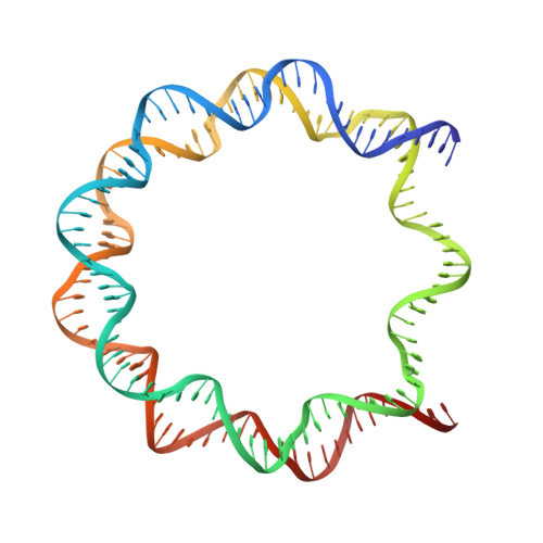
    6V2K

  • PubMed Abstract: 

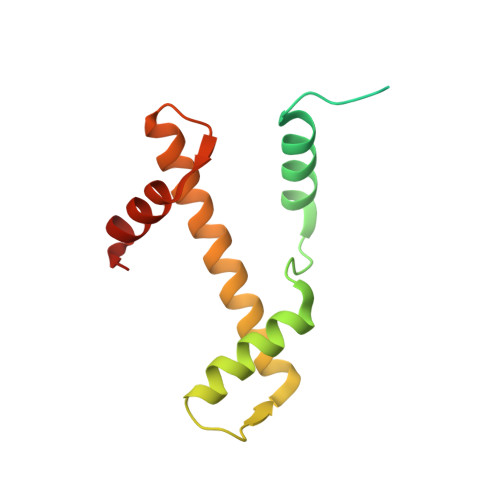
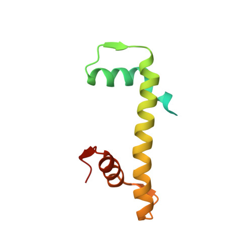
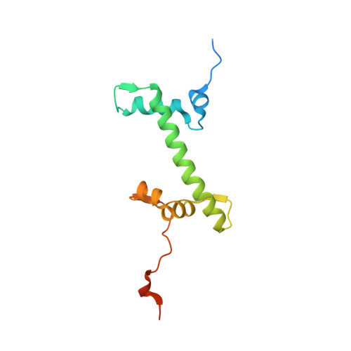
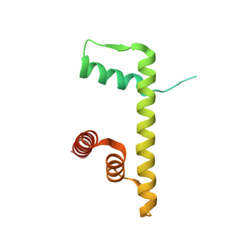
    H2A.B is an evolutionarily distant histone H2A variant that accumulates on DNA repair sites, DNA replication sites, and actively transcribing regions in genomes. In cells, H2A.B exchanges rapidly in chromatin, but the mechanism has remained enigmatic. In the present study, we found that the H2A.B-H2B dimer incorporated within the nucleosome exchanges with the canonical H2A-H2B dimer without assistance from additional factors, such as histone chaperones and nucleosome remodelers. High-speed atomic force microscopy revealed that the H2A.B nucleosome, but not the canonical H2A nucleosome, transiently forms an intermediate "open conformation", in which two H2A.B-H2B dimers may be detached from the H3-H4 tetramer and bind to the DNA regions near the entry/exit sites. Mutational analyses revealed that the H2A.B C-terminal region is responsible for the adoption of the open conformation and the H2A.B-H2B exchange in the nucleosome. These findings provide mechanistic insights into the histone exchange of the H2A.B nucleosome.


  • Organizational Affiliation

    Laboratory of Chromatin Structure and Function, Institute for Quantitative Biosciences, The University of Tokyo, Bunkyo-ku, Tokyo, Japan.


Macromolecules

Find similar proteins by:  (by identity cutoff)  |  3D Structure
Entity ID: 1
MoleculeChains Sequence LengthOrganismDetailsImage
Histone H3.1
A, E
139Homo sapiensMutation(s): 0 
Gene Names: 
UniProt & NIH Common Fund Data Resources
Find proteins for P68431 (Homo sapiens)
Explore P68431 
Go to UniProtKB:  P68431
PHAROS:  P68431
Entity Groups  
Sequence Clusters30% Identity50% Identity70% Identity90% Identity95% Identity100% Identity
UniProt GroupP68431
Sequence Annotations
Expand
  • Reference Sequence
Find similar proteins by:  (by identity cutoff)  |  3D Structure
Entity ID: 2
MoleculeChains Sequence LengthOrganismDetailsImage
Histone H4
B, F
106Homo sapiensMutation(s): 0 
Gene Names: 
UniProt & NIH Common Fund Data Resources
Find proteins for P62805 (Homo sapiens)
Explore P62805 
Go to UniProtKB:  P62805
PHAROS:  P62805
Entity Groups  
Sequence Clusters30% Identity50% Identity70% Identity90% Identity95% Identity100% Identity
UniProt GroupP62805
Sequence Annotations
Expand
  • Reference Sequence
Find similar proteins by:  (by identity cutoff)  |  3D Structure
Entity ID: 3
MoleculeChains Sequence LengthOrganismDetailsImage
Histone H2A
C, G
133Homo sapiensMutation(s): 0 
Gene Names: HIST1H2ABHIST1H2AEhCG_1640984hCG_1787383
UniProt & NIH Common Fund Data Resources
Find proteins for P04908 (Homo sapiens)
Explore P04908 
Go to UniProtKB:  P04908
PHAROS:  P04908
Entity Groups  
Sequence Clusters30% Identity50% Identity70% Identity90% Identity95% Identity100% Identity
UniProt GroupP04908
Sequence Annotations
Expand
  • Reference Sequence
Find similar proteins by:  (by identity cutoff)  |  3D Structure
Entity ID: 4
MoleculeChains Sequence LengthOrganismDetailsImage
Histone H2B type 1-J
D, H
129Homo sapiensMutation(s): 0 
Gene Names: HIST1H2BJH2BFR
UniProt & NIH Common Fund Data Resources
Find proteins for P06899 (Homo sapiens)
Explore P06899 
Go to UniProtKB:  P06899
PHAROS:  P06899
GTEx:  ENSG00000124635 
Entity Groups  
Sequence Clusters30% Identity50% Identity70% Identity90% Identity95% Identity100% Identity
UniProt GroupP06899
Sequence Annotations
Expand
  • Reference Sequence
Find similar nucleic acids by:  (by identity cutoff)  |  3D Structure
Entity ID: 5
MoleculeChains LengthOrganismImage
DNA (146-MER)
I, J
146Homo sapiens
Sequence Annotations
Expand
  • Reference Sequence
Small Molecules
Ligands 2 Unique
IDChains Name / Formula / InChI Key2D Diagram3D Interactions
MN
Query on MN

Download Ideal Coordinates CCD File 
M [auth E]
P [auth I]
Q [auth I]
R [auth I]
S [auth I]
M [auth E],
P [auth I],
Q [auth I],
R [auth I],
S [auth I],
T [auth I],
U [auth J],
V [auth J],
W [auth J],
X [auth J]
MANGANESE (II) ION
Mn
WAEMQWOKJMHJLA-UHFFFAOYSA-N
CL
Query on CL

Download Ideal Coordinates CCD File 
K [auth A],
L [auth C],
N [auth E],
O [auth G]
CHLORIDE ION
Cl
VEXZGXHMUGYJMC-UHFFFAOYSA-M
Experimental Data & Validation

Experimental Data

  • Method: X-RAY DIFFRACTION
  • Resolution: 2.60 Å
  • R-Value Free: 0.248 
  • R-Value Work: 0.197 
  • R-Value Observed: 0.199 
  • Space Group: P 21 21 21
Unit Cell:
Length ( Å )Angle ( ˚ )
a = 98.561α = 90
b = 107.711β = 90
c = 168.158γ = 90
Software Package:
Software NamePurpose
PHENIXrefinement
HKL-2000data reduction
HKL-2000data scaling
PDB_EXTRACTdata extraction
PHASERphasing

Structure Validation

View Full Validation Report

Currently 6V2K does not have a validation slider image.


Entry History & Funding Information

Deposition Data


Funding OrganizationLocationGrant Number
Japan Society for the Promotion of Science (JSPS)JapanJP18H05534
Japan Society for the Promotion of Science (JSPS)JapanJP17H01408
Japan Science and TechnologyJapanJPMJCR16G1
Japan Science and TechnologyJapanJPMJER1901
Japan Agency for Medical Research and Development (AMED)JapanJP19am0101076

Revision History  (Full details and data files)

  • Version 1.0: 2020-11-25
    Type: Initial release
  • Version 1.1: 2021-02-24
    Changes: Database references
  • Version 1.2: 2023-10-11
    Changes: Data collection, Database references, Refinement description