6USA

Crystal structure of tryptophan synthase from M. tuberculosis - aminoacrylate- and GSK1-bound form


Experimental Data Snapshot

  • Method: X-RAY DIFFRACTION
  • Resolution: 2.41 Å
  • R-Value Free: 0.192 
  • R-Value Work: 0.158 
  • R-Value Observed: 0.158 

wwPDB Validation   3D Report Full Report


Ligand Structure Quality Assessment 


This is version 1.3 of the entry. See complete history


Literature

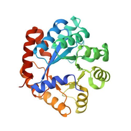
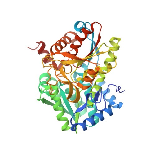
Allosteric inhibitors of Mycobacterium tuberculosis tryptophan synthase.

Michalska, K.Chang, C.Maltseva, N.I.Jedrzejczak, R.Robertson, G.T.Gusovsky, F.McCarren, P.Schreiber, S.L.Nag, P.P.Joachimiak, A.

(2020) Protein Sci 29: 779-788

  • DOI: https://doi.org/10.1002/pro.3825
  • Primary Citation of Related Structures:  
    6U6C, 6USA

  • PubMed Abstract: 

    Global dispersion of multidrug resistant bacteria is very common and evolution of antibiotic-resistance is occurring at an alarming rate, presenting a formidable challenge for humanity. The development of new therapeuthics with novel molecular targets is urgently needed. Current drugs primarily affect protein, nucleic acid, and cell wall synthesis. Metabolic pathways, including those involved in amino acid biosynthesis, have recently sparked interest in the drug discovery community as potential reservoirs of such novel targets. Tryptophan biosynthesis, utilized by bacteria but absent in humans, represents one of the currently studied processes with a therapeutic focus. It has been shown that tryptophan synthase (TrpAB) is required for survival of Mycobacterium tuberculosis in macrophages and for evading host defense, and therefore is a promising drug target. Here we present crystal structures of TrpAB with two allosteric inhibitors of M. tuberculosis tryptophan synthase that belong to sulfolane and indole-5-sulfonamide chemical scaffolds. We compare our results with previously reported structural and biochemical studies of another, azetidine-containing M. tuberculosis tryptophan synthase inhibitor. This work shows how structurally distinct ligands can occupy the same allosteric site and make specific interactions. It also highlights the potential benefit of targeting more variable allosteric sites of important metabolic enzymes.


  • Organizational Affiliation

    Center for Structural Genomics of Infectious Diseases, Consortium for Advanced Science and Engineering, University of Chicago, Chicago, Illinois.


Macromolecules
Find similar proteins by:  (by identity cutoff)  |  3D Structure
Entity ID: 1
MoleculeChains Sequence LengthOrganismDetailsImage
Tryptophan synthase alpha chain
A, C, E, G
276Mycobacterium tuberculosis H37RvMutation(s): 0 
Gene Names: trpARv1613MTCY01B2.05
EC: 4.2.1.20
UniProt
Find proteins for P9WFY1 (Mycobacterium tuberculosis (strain ATCC 25618 / H37Rv))
Explore P9WFY1 
Go to UniProtKB:  P9WFY1
Entity Groups  
Sequence Clusters30% Identity50% Identity70% Identity90% Identity95% Identity100% Identity
UniProt GroupP9WFY1
Sequence Annotations
Expand
  • Reference Sequence
Find similar proteins by:  (by identity cutoff)  |  3D Structure
Entity ID: 2
MoleculeChains Sequence LengthOrganismDetailsImage
Tryptophan synthase beta chain
B, D, F, H
410Mycobacterium tuberculosis H37RvMutation(s): 0 
Gene Names: trpBRv1612MTCY01B2.04
EC: 4.2.1.20
UniProt
Find proteins for P9WFX9 (Mycobacterium tuberculosis (strain ATCC 25618 / H37Rv))
Explore P9WFX9 
Go to UniProtKB:  P9WFX9
Entity Groups  
Sequence Clusters30% Identity50% Identity70% Identity90% Identity95% Identity100% Identity
UniProt GroupP9WFX9
Sequence Annotations
Expand
  • Reference Sequence
Small Molecules
Ligands 10 Unique
IDChains Name / Formula / InChI Key2D Diagram3D Interactions
PZJ (Subject of Investigation/LOI)
Query on PZJ

Download Ideal Coordinates CCD File 
L [auth B],
MB [auth H],
QA [auth F],
Y [auth D]
(3R,4R)-4-[4-(2-Chlorophenyl)piperazin-1-yl]-1,1-dioxothiolan-3-ol
C14 H19 Cl N2 O3 S
CRYVRLLTCKZRFB-KBPBESRZSA-N
P1T
Query on P1T

Download Ideal Coordinates CCD File 
M [auth B],
NB [auth H],
RA [auth F],
Z [auth D]
2-[({3-HYDROXY-2-METHYL-5-[(PHOSPHONOOXY)METHYL]PYRIDIN-4-YL}METHYL)AMINO]ACRYLIC ACID
C11 H15 N2 O7 P
BXUDKFHCAMQSRX-UHFFFAOYSA-N
PEG
Query on PEG

Download Ideal Coordinates CCD File 
V [auth C]DI(HYDROXYETHYL)ETHER
C4 H10 O3
MTHSVFCYNBDYFN-UHFFFAOYSA-N
MLI
Query on MLI

Download Ideal Coordinates CCD File 
DB [auth F]
K [auth A]
KA [auth D]
KB [auth G]
PA [auth E]
DB [auth F],
K [auth A],
KA [auth D],
KB [auth G],
PA [auth E],
X [auth C]
MALONATE ION
C3 H2 O4
OFOBLEOULBTSOW-UHFFFAOYSA-L
SO4
Query on SO4

Download Ideal Coordinates CCD File 
VB [auth H]SULFATE ION
O4 S
QAOWNCQODCNURD-UHFFFAOYSA-L
EDO
Query on EDO

Download Ideal Coordinates CCD File 
EB [auth F]
FB [auth F]
GB [auth F]
HB [auth F]
LA [auth D]
EB [auth F],
FB [auth F],
GB [auth F],
HB [auth F],
LA [auth D],
LB [auth G],
MA [auth D],
T [auth B],
U [auth B]
1,2-ETHANEDIOL
C2 H6 O2
LYCAIKOWRPUZTN-UHFFFAOYSA-N
ACT
Query on ACT

Download Ideal Coordinates CCD File 
CA [auth D],
GA [auth D],
VA [auth F]
ACETATE ION
C2 H3 O2
QTBSBXVTEAMEQO-UHFFFAOYSA-M
FMT
Query on FMT

Download Ideal Coordinates CCD File 
AA [auth D]
BA [auth D]
DA [auth D]
EA [auth D]
FA [auth D]
AA [auth D],
BA [auth D],
DA [auth D],
EA [auth D],
FA [auth D],
I [auth A],
IB [auth G],
J [auth A],
N [auth B],
NA [auth E],
O [auth B],
OA [auth E],
OB [auth H],
P [auth B],
PB [auth H],
Q [auth B],
QB [auth H],
R [auth B],
RB [auth H],
SA [auth F],
SB [auth H],
TA [auth F],
TB [auth H],
UA [auth F],
W [auth C],
WA [auth F],
XA [auth F],
YA [auth F]
FORMIC ACID
C H2 O2
BDAGIHXWWSANSR-UHFFFAOYSA-N
K
Query on K

Download Ideal Coordinates CCD File 
AB [auth F]
BB [auth F]
CB [auth F]
HA [auth D]
IA [auth D]
AB [auth F],
BB [auth F],
CB [auth F],
HA [auth D],
IA [auth D],
JA [auth D],
JB [auth G],
S [auth B],
UB [auth H],
ZA [auth F]
POTASSIUM ION
K
NPYPAHLBTDXSSS-UHFFFAOYSA-N
NA
Query on NA

Download Ideal Coordinates CCD File 
WB [auth H],
XB [auth H]
SODIUM ION
Na
FKNQFGJONOIPTF-UHFFFAOYSA-N
Experimental Data & Validation

Experimental Data

  • Method: X-RAY DIFFRACTION
  • Resolution: 2.41 Å
  • R-Value Free: 0.192 
  • R-Value Work: 0.158 
  • R-Value Observed: 0.158 
  • Space Group: P 21 21 21
Unit Cell:
Length ( Å )Angle ( ˚ )
a = 134.92α = 90
b = 160.037β = 90
c = 165.151γ = 90
Software Package:
Software NamePurpose
PHENIXrefinement
SCALEPACKdata scaling
PDB_EXTRACTdata extraction
HKL-3000data reduction
HKL-3000phasing

Structure Validation

View Full Validation Report


Ligand Structure Quality Assessment 


Entry History & Funding Information

Deposition Data


Funding OrganizationLocationGrant Number
Global Health Innovative Technology FundUnited States--

Revision History  (Full details and data files)

  • Version 1.0: 2020-09-30
    Type: Initial release
  • Version 1.1: 2022-08-31
    Changes: Advisory, Database references, Structure summary
  • Version 1.2: 2023-02-01
    Changes: Advisory
  • Version 1.3: 2023-10-25
    Changes: Data collection, Refinement description