6UO6

Crystal Structure of the R422Q missense variant of human PGM1


Experimental Data Snapshot

  • Method: X-RAY DIFFRACTION
  • Resolution: 2.15 Å
  • R-Value Free: 0.213 
  • R-Value Work: 0.175 
  • R-Value Observed: 0.177 

wwPDB Validation   3D Report Full Report

Currently 6UO6 does not have a validation slider image.


This is version 1.2 of the entry. See complete history


Literature

A missense variant remote from the active site impairs stability of human phosphoglucomutase 1.

Stiers, K.M.Hansen, R.P.Daghlas, B.A.Mason, K.N.Zhu, J.S.Jakeman, D.L.Beamer, L.J.

(2020) J Inherit Metab Dis 43: 861-870

  • DOI: https://doi.org/10.1002/jimd.12222
  • Primary Citation of Related Structures:  
    6UO6

  • PubMed Abstract: 

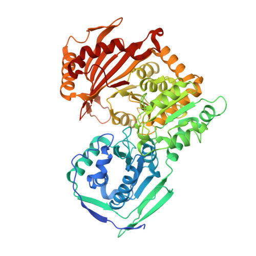
    Missense variants of human phosphoglucomutase 1 (PGM1) cause the inherited metabolic disease known as PGM1 deficiency. This condition is categorised as both a glycogen storage disease and a congenital disorder of glycosylation. Approximately 20 missense variants of PGM1 are linked to PGM1 deficiency, and biochemical studies have suggested that they fall into two general categories: those affecting the active site and catalytic efficiency, and those that appear to impair protein folding and/or stability. In this study, we characterise a novel variant of Arg422, a residue distal from the active site of PGM1 and the site of a previously identified disease-related variant (Arg422Trp). In prior studies, the R422W variant was found to produce insoluble protein in a recombinant expression system, precluding further in vitro characterisation. Here we investigate an alternative variant of this residue, Arg422Gln, which is amenable to experimental characterisation presumably due to its more conservative physicochemical substitution. Biochemical, crystallographic, and computational studies of R422Q establish that this variant causes only minor changes in catalytic efficiency and 3D structure, but is nonetheless dramatically reduced in stability. Unexpectedly, binding of a substrate analog is found to further destabilise the protein, in contrast to its stabilising effect on wild-type PGM1 and several other missense variants. This work establishes Arg422 as a lynchpin residue for the stability of PGM1 and supports the impairment of protein stability as a pathomechanism for variants that cause PGM1 deficiency. SYNOPSIS: Biochemical and structural studies of a missense variant far from the active site of human PGM1 identify a residue with a key role in enzyme stability.


  • Organizational Affiliation

    Department of Biochemistry, University of Missouri, Columbia, Missouri, USA.


Macromolecules
Find similar proteins by:  (by identity cutoff)  |  3D Structure
Entity ID: 1
MoleculeChains Sequence LengthOrganismDetailsImage
Phosphoglucomutase-1
A, B
585Homo sapiensMutation(s): 1 
Gene Names: PGM1
EC: 5.4.2.2
UniProt & NIH Common Fund Data Resources
Find proteins for P36871 (Homo sapiens)
Explore P36871 
Go to UniProtKB:  P36871
PHAROS:  P36871
GTEx:  ENSG00000079739 
Entity Groups  
Sequence Clusters30% Identity50% Identity70% Identity90% Identity95% Identity100% Identity
UniProt GroupP36871
Sequence Annotations
Expand
  • Reference Sequence
Small Molecules
Ligands 2 Unique
IDChains Name / Formula / InChI Key2D Diagram3D Interactions
SO4
Query on SO4

Download Ideal Coordinates CCD File 
D [auth A]
E [auth A]
F [auth A]
G [auth A]
H [auth A]
D [auth A],
E [auth A],
F [auth A],
G [auth A],
H [auth A],
I [auth A],
K [auth B],
L [auth B]
SULFATE ION
O4 S
QAOWNCQODCNURD-UHFFFAOYSA-L
MG
Query on MG

Download Ideal Coordinates CCD File 
C [auth A],
J [auth B]
MAGNESIUM ION
Mg
JLVVSXFLKOJNIY-UHFFFAOYSA-N
Experimental Data & Validation

Experimental Data

  • Method: X-RAY DIFFRACTION
  • Resolution: 2.15 Å
  • R-Value Free: 0.213 
  • R-Value Work: 0.175 
  • R-Value Observed: 0.177 
  • Space Group: P 41 21 2
Unit Cell:
Length ( Å )Angle ( ˚ )
a = 171.804α = 90
b = 171.804β = 90
c = 99.278γ = 90
Software Package:
Software NamePurpose
PHENIXrefinement
XDSdata reduction
Aimlessdata scaling
PHENIXphasing

Structure Validation

View Full Validation Report

Currently 6UO6 does not have a validation slider image.


Entry History & Funding Information

Deposition Data


Funding OrganizationLocationGrant Number
National Science Foundation (NSF, United States)United StatesMCB-1409898

Revision History  (Full details and data files)

  • Version 1.0: 2020-03-04
    Type: Initial release
  • Version 1.1: 2020-07-22
    Changes: Database references
  • Version 1.2: 2023-10-11
    Changes: Advisory, Data collection, Database references, Derived calculations, Refinement description