6TNP

Crystal structure of the ScFv-5E5 in complex with a Tn glycopeptide


Experimental Data Snapshot

  • Method: X-RAY DIFFRACTION
  • Resolution: 3.00 Å
  • R-Value Free: 0.251 
  • R-Value Work: 0.203 
  • R-Value Observed: 0.205 

wwPDB Validation   3D Report Full Report


Ligand Structure Quality Assessment 


This is version 1.3 of the entry. See complete history


Literature

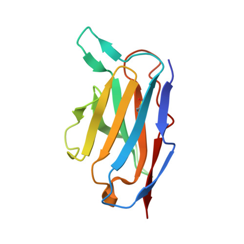
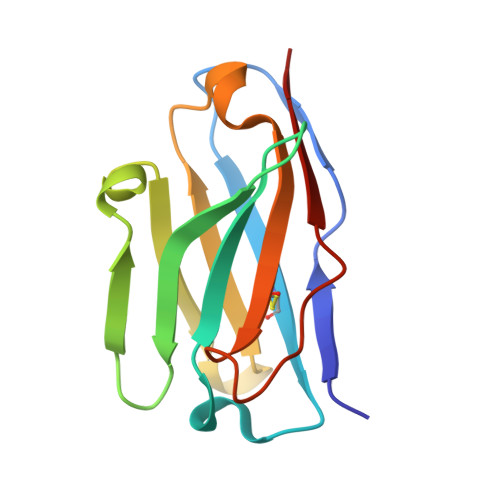
Structural characterization of an unprecedented lectin-like antitumoral anti-MUC1 antibody.

Macias-Leon, J.Bermejo, I.A.Asin, A.Garcia-Garcia, A.Companon, I.Jimenez-Moreno, E.Coelho, H.Mangini, V.Albuquerque, I.S.Marcelo, F.Asensio, J.L.Bernardes, G.J.L.Joshi, H.J.Fiammengo, R.Blixt, O.Hurtado-Guerrero, R.Corzana, F.

(2020) Chem Commun (Camb) 56: 15137-15140

  • DOI: https://doi.org/10.1039/d0cc06349e
  • Primary Citation of Related Structures:  
    6TNP

  • PubMed Abstract: 

    The molecular basis of antibody 5E5, which recognizes the entire GalNAc unit as a primary epitope is disclosed. The antibody's contacts with the peptide are mostly limited to two residues, allowing it to show some degree of promiscuity. These findings open the door to the chemical design of peptide-mimetics for developing efficient anti-cancer vaccines and diagnostic tools.


  • Organizational Affiliation

    Institute of Biocomputation and Physics of Complex Systems (BIFI), University of Zaragoza, Mariano Esquillor s/n, Campus Rio Ebro, Edificio I+D, Zaragoza, Spain. rhurtado@bifi.es.


Macromolecules
Find similar proteins by:  (by identity cutoff)  |  3D Structure
Entity ID: 1
MoleculeChains Sequence LengthOrganismDetailsImage
Light chain of our ScFv-5E5
A, C, E, G, J
A, C, E, G, J, L
113Mus musculusMutation(s): 0 
Entity Groups  
Sequence Clusters30% Identity50% Identity70% Identity90% Identity95% Identity100% Identity
Sequence Annotations
Expand
  • Reference Sequence
Find similar proteins by:  (by identity cutoff)  |  3D Structure
Entity ID: 2
MoleculeChains Sequence LengthOrganismDetailsImage
Heavy chain of our ScFv-5E5
B, D, F, H, I
B, D, F, H, I, K
116Mus musculusMutation(s): 0 
Entity Groups  
Sequence Clusters30% Identity50% Identity70% Identity90% Identity95% Identity100% Identity
Sequence Annotations
Expand
  • Reference Sequence
Small Molecules
Ligands 1 Unique
IDChains Name / Formula / InChI Key2D Diagram3D Interactions
NO8 (Subject of Investigation/LOI)
Query on NO8

Download Ideal Coordinates CCD File 
M [auth B],
N [auth D],
O [auth F],
P [auth H],
Q [auth I]
(2~{S})-~{N}-[2-[[(2~{S})-1-[[(2~{S},3~{R})-3-[(2~{S},3~{R},4~{R},5~{R},6~{R})-3-acetamido-6-(hydroxymethyl)-4,5-bis(oxidanyl)oxan-2-yl]oxy-1-[[(2~{S})-1-[(2~{S})-2-aminocarbonylpyrrolidin-1-yl]-1-oxidanylidene-propan-2-yl]amino]-1-oxidanylidene-butan-2-yl]amino]-3-oxidanyl-1-oxidanylidene-propan-2-yl]amino]-2-oxidanylidene-ethyl]-1-[(2~{S})-2-azanylpropanoyl]pyrrolidine-2-carboxamide
C33 H55 N9 O14
SQMBDAVTEXMIPT-PEECNIKTSA-N
Experimental Data & Validation

Experimental Data

  • Method: X-RAY DIFFRACTION
  • Resolution: 3.00 Å
  • R-Value Free: 0.251 
  • R-Value Work: 0.203 
  • R-Value Observed: 0.205 
  • Space Group: P 21 21 21
Unit Cell:
Length ( Å )Angle ( ˚ )
a = 86.573α = 90
b = 113.195β = 90
c = 151.201γ = 90
Software Package:
Software NamePurpose
REFMACrefinement
XDSdata reduction
SCALAdata scaling
PHASERphasing

Structure Validation

View Full Validation Report


Ligand Structure Quality Assessment 


Entry History 

Deposition Data

Revision History  (Full details and data files)

  • Version 1.0: 2020-11-18
    Type: Initial release
  • Version 1.1: 2020-12-02
    Changes: Database references
  • Version 1.2: 2020-12-16
    Changes: Database references
  • Version 1.3: 2024-01-24
    Changes: Data collection, Database references, Refinement description