


Crystal structure of mouse PRMT6 in complex with inhibitors
Bonnefond, L., Cavarelli, J.To be published.
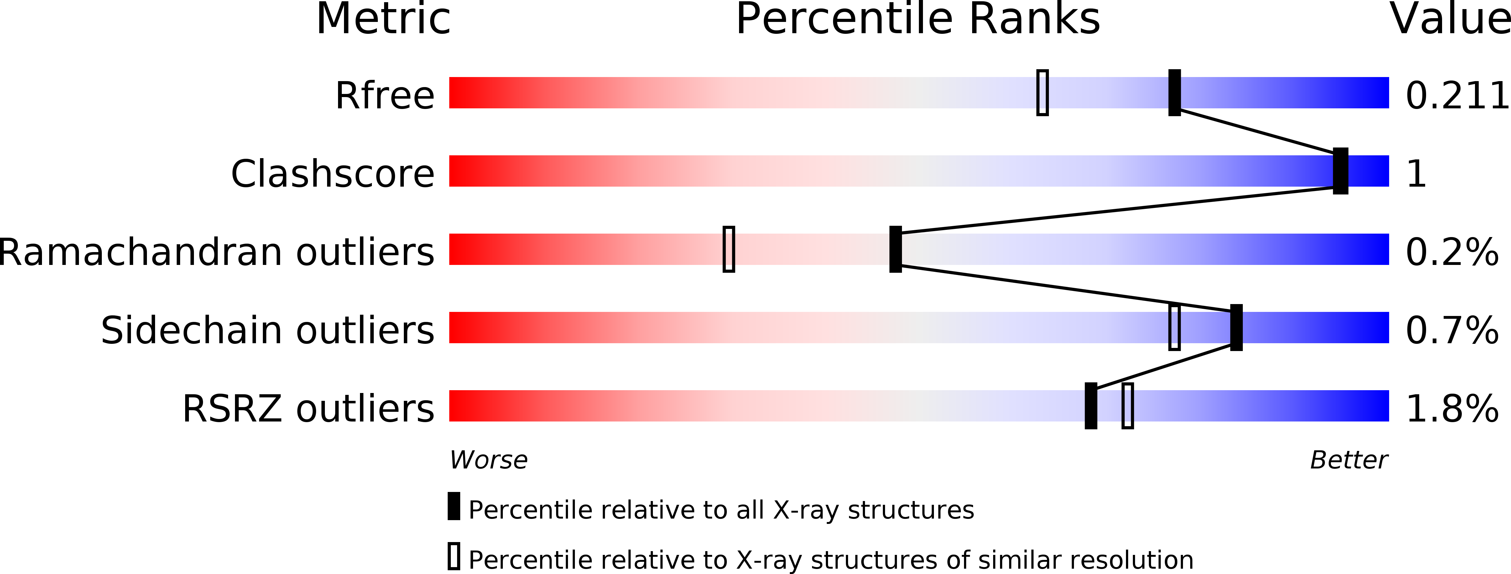
Experimental Data Snapshot
Entity ID: 1 | |||||
|---|---|---|---|---|---|
| Molecule | Chains | Sequence Length | Organism | Details | Image |
| Protein arginine N-methyltransferase 6 | 386 | Mus musculus | Mutation(s): 1 Gene Names: Prmt6, Hrmt1l6 EC: 2.1.1.319 | 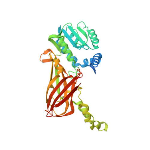 | |
UniProt & NIH Common Fund Data Resources | |||||
Find proteins for Q6NZB1 (Mus musculus) Explore Q6NZB1 Go to UniProtKB: Q6NZB1 | |||||
IMPC: MGI:2139971 | |||||
Entity Groups | |||||
| Sequence Clusters | 30% Identity50% Identity70% Identity90% Identity95% Identity100% Identity | ||||
| UniProt Group | Q6NZB1 | ||||
Sequence AnnotationsExpand | |||||
| |||||
| Ligands 2 Unique | |||||
|---|---|---|---|---|---|
| ID | Chains | Name / Formula / InChI Key | 2D Diagram | 3D Interactions | |
| GJV (Subject of Investigation/LOI) Query on GJV | D [auth A], F [auth B] | 9-(7-{[amino(iminio)methyl]amino}-5,6,7-trideoxy-beta-D-ribo-heptofuranosyl)-9H-purin-6-amine C13 H21 N8 O3 NGQAXJZDWQDSQY-WOUKDFQISA-O |  | ||
| IOD Query on IOD | C [auth A], E [auth B] | IODIDE ION I XMBWDFGMSWQBCA-UHFFFAOYSA-M |  | ||
| Length ( Å ) | Angle ( ˚ ) |
|---|---|
| a = 41.95 | α = 90 |
| b = 118.101 | β = 103.45 |
| c = 72.093 | γ = 90 |
| Software Name | Purpose |
|---|---|
| XDS | data reduction |
| Aimless | data scaling |
| PHENIX | refinement |
| PDB_EXTRACT | data extraction |
| AutoSol | phasing |
RCSB PDB (citation) is hosted by
RCSB PDB is a member of the

