6SMO

AntDE:AntF (apo): type II PKS acyl-carrier protein in complex with its ketosynthase bound to the hexaketide


Experimental Data Snapshot

  • Method: X-RAY DIFFRACTION
  • Resolution: 2.70 Å
  • R-Value Free: 0.265 
  • R-Value Work: 0.226 
  • R-Value Observed: 0.229 

wwPDB Validation   3D Report Full Report


Ligand Structure Quality Assessment 


This is version 1.4 of the entry. See complete history


Literature

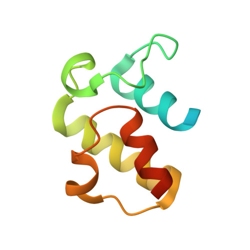
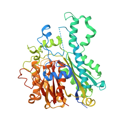
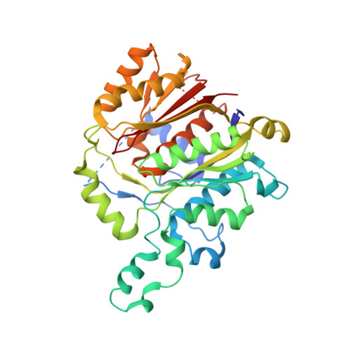
Structural snapshots of the minimal PKS system responsible for octaketide biosynthesis.

Brauer, A.Zhou, Q.Grammbitter, G.L.C.Schmalhofer, M.Ruhl, M.Kaila, V.R.I.Bode, H.B.Groll, M.

(2020) Nat Chem 12: 755-763

  • DOI: https://doi.org/10.1038/s41557-020-0491-7
  • Primary Citation of Related Structures:  
    6SM4, 6SM6, 6SMD, 6SMO, 6SMP

  • PubMed Abstract: 

    Type II polyketide synthases (PKSs) are multi-enzyme complexes that produce secondary metabolites of medical relevance. Chemical backbones of such polyketides are produced by minimal PKS systems that consist of a malonyl transacylase, an acyl carrier protein and an α/β heterodimeric ketosynthase. Here, we present X-ray structures of all ternary complexes that constitute the minimal PKS system for anthraquinone biosynthesis in Photorhabdus luminescens. In addition, we characterize this invariable core using molecular simulations, mutagenesis experiments and functional assays. We show that malonylation of the acyl carrier protein is accompanied by major structural rearrangements in the transacylase. Principles of an ongoing chain elongation are derived from the ternary complex with a hexaketide covalently linking the heterodimeric ketosynthase with the acyl carrier protein. Our results for the minimal PKS system provide mechanistic understanding of PKSs and a fundamental basis for engineering PKS pathways for future applications.


  • Organizational Affiliation

    Center for Integrated Protein Science Munich (CIPSM), Department of Chemistry, Technische Universität München, Garching, Germany.


Macromolecules
Find similar proteins by:  (by identity cutoff)  |  3D Structure
Entity ID: 1
MoleculeChains Sequence LengthOrganismDetailsImage
Acyl carrier protein97Photorhabdus luminescensMutation(s): 0 
Gene Names: C6H68_21855
UniProt
Find proteins for Q7MZT5 (Photorhabdus laumondii subsp. laumondii (strain DSM 15139 / CIP 105565 / TT01))
Explore Q7MZT5 
Go to UniProtKB:  Q7MZT5
Entity Groups  
Sequence Clusters30% Identity50% Identity70% Identity90% Identity95% Identity100% Identity
UniProt GroupQ7MZT5
Sequence Annotations
Expand
  • Reference Sequence
Find similar proteins by:  (by identity cutoff)  |  3D Structure
Entity ID: 2
MoleculeChains Sequence LengthOrganismDetailsImage
PKS_KS domain-containing protein
B, D, F
443Photorhabdus laumondii subsp. laumondii TTO1Mutation(s): 0 
Gene Names: plu4191
UniProt
Find proteins for Q7MZT3 (Photorhabdus laumondii subsp. laumondii (strain DSM 15139 / CIP 105565 / TT01))
Explore Q7MZT3 
Go to UniProtKB:  Q7MZT3
Entity Groups  
Sequence Clusters30% Identity50% Identity70% Identity90% Identity95% Identity100% Identity
UniProt GroupQ7MZT3
Sequence Annotations
Expand
  • Reference Sequence
Find similar proteins by:  (by identity cutoff)  |  3D Structure
Entity ID: 3
MoleculeChains Sequence LengthOrganismDetailsImage
Ketoacyl_synth_N domain-containing protein
C, E, G
371Photorhabdus laumondii subsp. laumondii TTO1Mutation(s): 0 
Gene Names: plu4190
UniProt
Find proteins for Q7MZT4 (Photorhabdus laumondii subsp. laumondii (strain DSM 15139 / CIP 105565 / TT01))
Explore Q7MZT4 
Go to UniProtKB:  Q7MZT4
Entity Groups  
Sequence Clusters30% Identity50% Identity70% Identity90% Identity95% Identity100% Identity
UniProt GroupQ7MZT4
Sequence Annotations
Expand
  • Reference Sequence
Experimental Data & Validation

Experimental Data

  • Method: X-RAY DIFFRACTION
  • Resolution: 2.70 Å
  • R-Value Free: 0.265 
  • R-Value Work: 0.226 
  • R-Value Observed: 0.229 
  • Space Group: P 41
Unit Cell:
Length ( Å )Angle ( ˚ )
a = 107.65α = 90
b = 107.65β = 90
c = 201.33γ = 90
Software Package:
Software NamePurpose
REFMACrefinement
PDB_EXTRACTdata extraction
XDSdata reduction
XSCALEdata scaling
PHASERphasing

Structure Validation

View Full Validation Report


Ligand Structure Quality Assessment 


Entry History 

Deposition Data

Revision History  (Full details and data files)

  • Version 1.0: 2020-05-27
    Type: Initial release
  • Version 1.1: 2020-07-15
    Changes: Database references
  • Version 1.2: 2020-08-05
    Changes: Database references
  • Version 1.3: 2020-11-11
    Changes: Derived calculations
  • Version 1.4: 2024-01-24
    Changes: Data collection, Database references, Refinement description