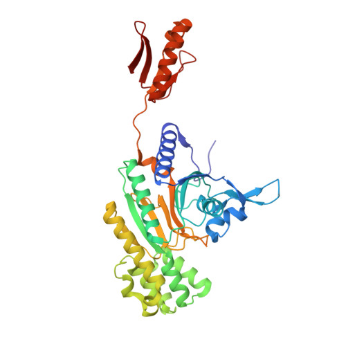
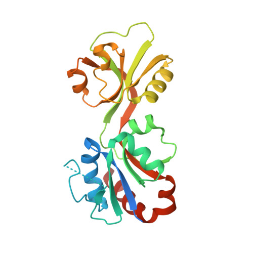
6R02

Psychrobacter arcticus ATP phosphoribosyltransferase bound to histidine and PRPP


Experimental Data Snapshot

  • Method: X-RAY DIFFRACTION
  • Resolution: 2.65 Å
  • R-Value Free: 0.270 
  • R-Value Work: 0.236 
  • R-Value Observed: 0.238 

wwPDB Validation   3D Report Full Report


Ligand Structure Quality Assessment 


This is version 1.2 of the entry. See complete history


Literature

Mapping the Structural Path for Allosteric Inhibition of a Short-Form ATP Phosphoribosyltransferase by Histidine.

Thomson, C.M.Alphey, M.S.Fisher, G.da Silva, R.G.

(2019) Biochemistry 58: 3078-3086

  • DOI: https://doi.org/10.1021/acs.biochem.9b00282
  • Primary Citation of Related Structures:  
    6R02

  • PubMed Abstract: 

    ATP phosphoribosyltransferase (ATPPRT) catalyzes the first step of histidine biosynthesis, being allosterically inhibited by the final product of the pathway. Allosteric inhibition of long-form ATPPRTs by histidine has been extensively studied, but inhibition of short-form ATPPRTs is poorly understood. Short-form ATPPRTs are hetero-octamers formed by four catalytic subunits (HisG S ) and four regulatory subunits (HisZ). HisG S alone is catalytically active and insensitive to histidine. HisZ enhances catalysis by HisG S in the absence of histidine but mediates allosteric inhibition in its presence. Here, steady-state and pre-steady-state kinetics establish that histidine is a noncompetitive inhibitor of short-form ATPPRT and that inhibition does not occur by dissociating HisG S from the hetero-octamer. The crystal structure of ATPPRT in complex with histidine and the substrate 5-phospho-α-d-ribosyl-1-pyrophosphate was determined, showing histidine bound solely to HisZ, with four histidine molecules per hetero-octamer. Histidine binding involves the repositioning of two HisZ loops. The histidine-binding loop moves closer to histidine to establish polar contacts. This leads to a hydrogen bond between its Tyr263 and His104 in the Asp101-Leu117 loop. The Asp101-Leu117 loop leads to the HisZ-HisG S interface, and in the absence of histidine, its motion prompts HisG S conformational changes responsible for catalytic activation. Following histidine binding, interaction with the histidine-binding loop may prevent the Asp101-Leu117 loop from efficiently sampling conformations conducive to catalytic activation. Tyr263Phe- Pa HisZ-containing Pa ATPPRT, however, is less susceptible though not insensitive to histidine inhibition, suggesting the Tyr263-His104 interaction may be relevant to yet not solely responsible for transmission of the allosteric signal.


  • Organizational Affiliation

    School of Biology, Biomedical Sciences Research Complex , University of St Andrews , St Andrews , Fife KY16 9ST , U.K.


Macromolecules
Find similar proteins by:  (by identity cutoff)  |  3D Structure
Entity ID: 1
MoleculeChains Sequence LengthOrganismDetailsImage
ATP phosphoribosyltransferase regulatory subunit
A, B, C, D
388Psychrobacter arcticusMutation(s): 0 
Gene Names: hisZPsyc_0676
UniProt
Find proteins for Q4FTX3 (Psychrobacter arcticus (strain DSM 17307 / VKM B-2377 / 273-4))
Explore Q4FTX3 
Go to UniProtKB:  Q4FTX3
Entity Groups  
Sequence Clusters30% Identity50% Identity70% Identity90% Identity95% Identity100% Identity
UniProt GroupQ4FTX3
Sequence Annotations
Expand
  • Reference Sequence
Find similar proteins by:  (by identity cutoff)  |  3D Structure
Entity ID: 2
MoleculeChains Sequence LengthOrganismDetailsImage
ATP phosphoribosyltransferase
E, F, G, H
232Psychrobacter arcticusMutation(s): 0 
Gene Names: hisGPsyc_1903
EC: 2.4.2.17
UniProt
Find proteins for Q4FQF7 (Psychrobacter arcticus (strain DSM 17307 / VKM B-2377 / 273-4))
Explore Q4FQF7 
Go to UniProtKB:  Q4FQF7
Entity Groups  
Sequence Clusters30% Identity50% Identity70% Identity90% Identity95% Identity100% Identity
UniProt GroupQ4FQF7
Sequence Annotations
Expand
  • Reference Sequence
Experimental Data & Validation

Experimental Data

  • Method: X-RAY DIFFRACTION
  • Resolution: 2.65 Å
  • R-Value Free: 0.270 
  • R-Value Work: 0.236 
  • R-Value Observed: 0.238 
  • Space Group: P 1 21 1
Unit Cell:
Length ( Å )Angle ( ˚ )
a = 93.253α = 90
b = 147.31β = 103.11
c = 99.327γ = 90
Software Package:
Software NamePurpose
REFMACrefinement
XDSdata reduction
Aimlessdata scaling
MOLREPphasing
PDB_EXTRACTdata extraction

Structure Validation

View Full Validation Report


Ligand Structure Quality Assessment 


Entry History 

Deposition Data

Revision History  (Full details and data files)

  • Version 1.0: 2019-08-07
    Type: Initial release
  • Version 1.1: 2020-07-29
    Type: Remediation
    Reason: Carbohydrate remediation
    Changes: Data collection, Derived calculations, Structure summary
  • Version 1.2: 2024-01-24
    Changes: Data collection, Database references, Refinement description, Structure summary