


Crystal structure of human Methylmalonyl-CoA epimerase (MCEE) p.Arg143Cys variant
Bailey, H.J., Chaikuid, A., Krysztofinska, E., Froese, D.S., Sorrell, F.J., Diaz-Saez, L., Kennedy, E., Edwards, A.M., Bountra, C., Yue, W.W.To be published.
Experimental Data Snapshot
wwPDB Validation 3D Report Full Report

Entity ID: 1 | |||||
|---|---|---|---|---|---|
| Molecule | Chains | Sequence Length | Organism | Details | Image |
| Methylmalonyl-CoA epimerase, mitochondrial | A [auth C], B [auth A], C [auth B], D | 155 | Homo sapiens | Mutation(s): 2 Gene Names: MCEE EC: 5.1.99.1 | 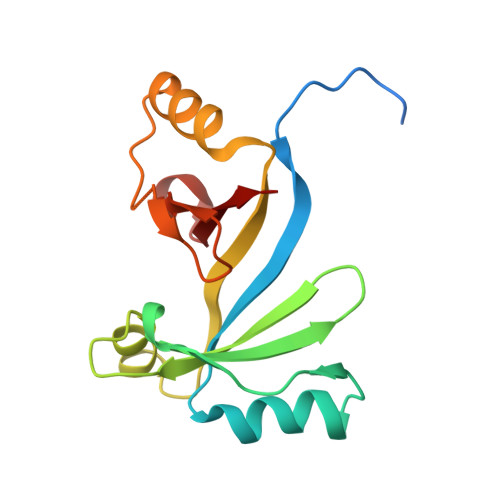 |
UniProt & NIH Common Fund Data Resources | |||||
Find proteins for Q96PE7 (Homo sapiens) Explore Q96PE7 Go to UniProtKB: Q96PE7 | |||||
PHAROS: Q96PE7 GTEx: ENSG00000124370 | |||||
Entity Groups | |||||
| Sequence Clusters | 30% Identity50% Identity70% Identity90% Identity95% Identity100% Identity | ||||
| UniProt Group | Q96PE7 | ||||
Sequence AnnotationsExpand | |||||
| |||||
| Ligands 1 Unique | |||||
|---|---|---|---|---|---|
| ID | Chains | Name / Formula / InChI Key | 2D Diagram | 3D Interactions | |
| CO Query on CO | E [auth C], F [auth A], G [auth B], H [auth D] | COBALT (II) ION Co XLJKHNWPARRRJB-UHFFFAOYSA-N |  | ||
| Length ( Å ) | Angle ( ˚ ) |
|---|---|
| a = 53.162 | α = 90 |
| b = 66.99 | β = 90.02 |
| c = 77.125 | γ = 90 |
| Software Name | Purpose |
|---|---|
| PHENIX | refinement |
| PDB_EXTRACT | data extraction |
| XDS | data reduction |
| STARANISO | data scaling |
| PHASER | phasing |
RCSB PDB (citation) is hosted by
RCSB PDB is a member of the

