6Q6N

Crystal structure of recombinant human beta-glucocerebrosidase in complex with biphenyl-cyclophellitol inhibitor (ME655)


Experimental Data Snapshot

  • Method: X-RAY DIFFRACTION
  • Resolution: 1.63 Å
  • R-Value Free: 0.212 
  • R-Value Work: 0.182 
  • R-Value Observed: 0.183 

wwPDB Validation   3D Report Full Report


Ligand Structure Quality Assessment 


This is version 2.1 of the entry. See complete history


Literature

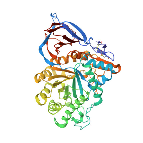
Functionalized Cyclophellitols Are Selective Glucocerebrosidase Inhibitors and Induce a Bona Fide Neuropathic Gaucher Model in Zebrafish.

Artola, M.Kuo, C.L.Lelieveld, L.T.Rowland, R.J.van der Marel, G.A.Codee, J.D.C.Boot, R.G.Davies, G.J.Aerts, J.M.F.G.Overkleeft, H.S.

(2019) J Am Chem Soc 141: 4214-4218

  • DOI: https://doi.org/10.1021/jacs.9b00056
  • Primary Citation of Related Structures:  
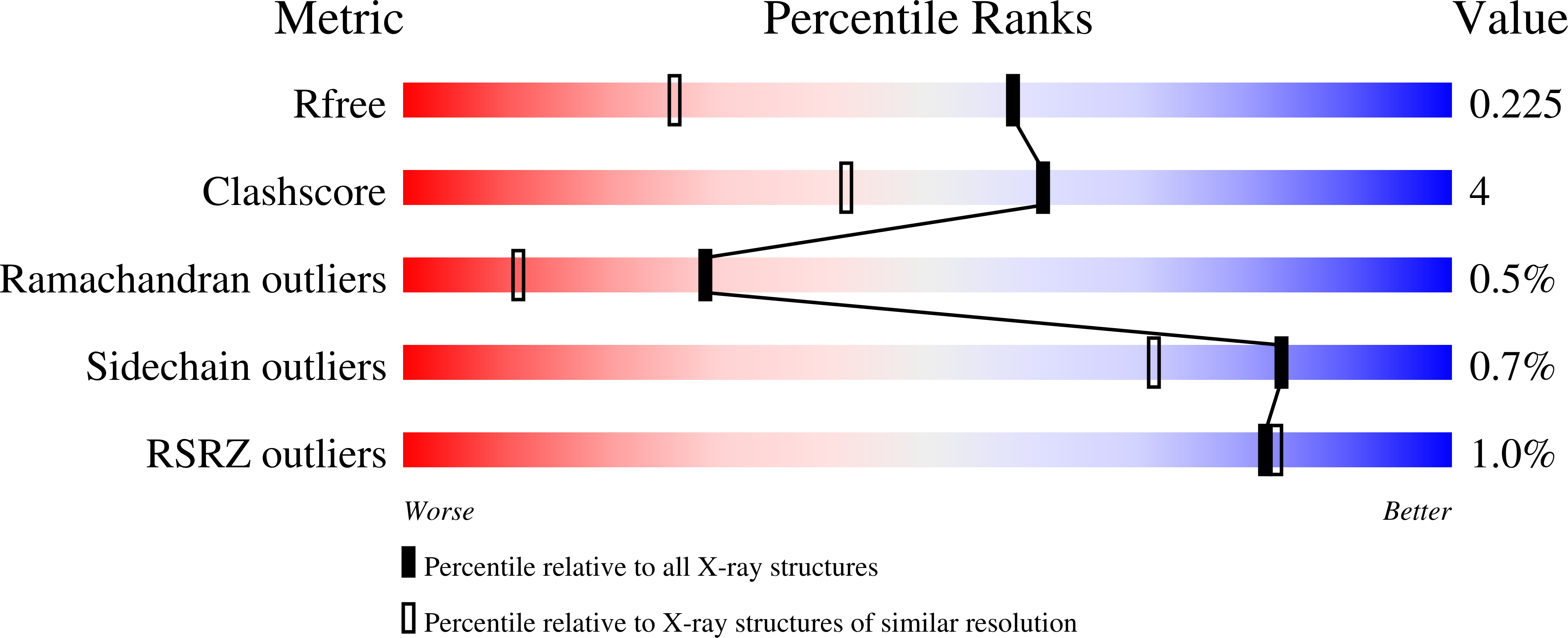
    6Q6K, 6Q6L, 6Q6N

  • PubMed Abstract: 

    Gaucher disease is caused by inherited deficiency in glucocerebrosidase (GBA, a retaining β-glucosidase), and deficiency in GBA constitutes the largest known genetic risk factor for Parkinson's disease. In the past, animal models of Gaucher disease have been generated by treatment with the mechanism-based GBA inhibitors, conduritol B epoxide (CBE), and cyclophellitol. Both compounds, however, also target other retaining glycosidases, rendering generation and interpretation of such chemical knockout models complicated. Here we demonstrate that cyclophellitol derivatives carrying a bulky hydrophobic substituent at C8 are potent and selective GBA inhibitors and that an unambiguous Gaucher animal model can be readily generated by treatment of zebrafish with these.


  • Organizational Affiliation

    Department of Chemistry, York Structural Biology Laboratory , University of York , Heslington, York YO10 5DD , United Kingdom.


Macromolecules
Find similar proteins by:  (by identity cutoff)  |  3D Structure
Entity ID: 1
MoleculeChains Sequence LengthOrganismDetailsImage
Glucosylceramidase
A, B
497Homo sapiensMutation(s): 1 
Gene Names: GBAGCGLUC
EC: 3.2.1.45
UniProt & NIH Common Fund Data Resources
Find proteins for P04062 (Homo sapiens)
Explore P04062 
Go to UniProtKB:  P04062
PHAROS:  P04062
GTEx:  ENSG00000177628 
Entity Groups  
Sequence Clusters30% Identity50% Identity70% Identity90% Identity95% Identity100% Identity
UniProt GroupP04062
Sequence Annotations
Expand
  • Reference Sequence
Oligosaccharides

Help

Entity ID: 2
MoleculeChains Length2D Diagram Glycosylation3D Interactions
2-acetamido-2-deoxy-beta-D-glucopyranose-(1-4)-2-acetamido-2-deoxy-beta-D-glucopyranose
C, D
2N-Glycosylation
Glycosylation Resources
GlyTouCan:  G42666HT
GlyCosmos:  G42666HT
GlyGen:  G42666HT
Small Molecules
Ligands 4 Unique
IDChains Name / Formula / InChI Key2D Diagram3D Interactions
HK2
Query on HK2

Download Ideal Coordinates CCD File 
RA [auth B],
V [auth A]
(1~{S},3~{S},4~{R},6~{R})-2,3,4,6-tetrakis(oxidanyl)-5-[[4-[3-(4-phenylphenoxy)propyl]-1,2,3-triazol-1-yl]methyl]cyclohexan-1-olate
C24 H28 N3 O6
IXNAMNDUIMARJW-WVYLNQBTSA-N
NAG
Query on NAG

Download Ideal Coordinates CCD File 
QA [auth B]2-acetamido-2-deoxy-beta-D-glucopyranose
C8 H15 N O6
OVRNDRQMDRJTHS-FMDGEEDCSA-N
SO4
Query on SO4

Download Ideal Coordinates CCD File 
BA [auth B]
CA [auth B]
DA [auth B]
E [auth A]
EA [auth B]
BA [auth B],
CA [auth B],
DA [auth B],
E [auth A],
EA [auth B],
F [auth A],
G [auth A],
H [auth A],
I [auth A],
K [auth A],
MA [auth B],
N [auth A],
R [auth A],
SA [auth B],
T [auth A]
SULFATE ION
O4 S
QAOWNCQODCNURD-UHFFFAOYSA-L
ACT
Query on ACT

Download Ideal Coordinates CCD File 
AA [auth A]
FA [auth B]
GA [auth B]
HA [auth B]
IA [auth B]
AA [auth A],
FA [auth B],
GA [auth B],
HA [auth B],
IA [auth B],
J [auth A],
JA [auth B],
KA [auth B],
L [auth A],
LA [auth B],
M [auth A],
NA [auth B],
O [auth A],
OA [auth B],
P [auth A],
PA [auth B],
Q [auth A],
S [auth A],
TA [auth B],
U [auth A],
W [auth A],
X [auth A],
Y [auth A],
Z [auth A]
ACETATE ION
C2 H3 O2
QTBSBXVTEAMEQO-UHFFFAOYSA-M
Experimental Data & Validation

Experimental Data

  • Method: X-RAY DIFFRACTION
  • Resolution: 1.63 Å
  • R-Value Free: 0.212 
  • R-Value Work: 0.182 
  • R-Value Observed: 0.183 
  • Space Group: C 2 2 21
Unit Cell:
Length ( Å )Angle ( ˚ )
a = 110.383α = 90
b = 285.152β = 90
c = 91.903γ = 90
Software Package:
Software NamePurpose
REFMACrefinement
xia2data reduction
Aimlessdata scaling
MOLREPphasing

Structure Validation

View Full Validation Report


Ligand Structure Quality Assessment 


Entry History & Funding Information

Deposition Data


Funding OrganizationLocationGrant Number
Biotechnology and Biological Sciences Research CouncilUnited KingdomBB/M011151/1

Revision History  (Full details and data files)

  • Version 1.0: 2019-03-27
    Type: Initial release
  • Version 1.1: 2019-05-08
    Changes: Data collection, Derived calculations
  • Version 1.2: 2019-07-10
    Changes: Data collection
  • Version 2.0: 2020-07-29
    Type: Remediation
    Reason: Carbohydrate remediation
    Changes: Advisory, Atomic model, Data collection, Derived calculations, Structure summary
  • Version 2.1: 2024-01-24
    Changes: Data collection, Database references, Refinement description, Structure summary