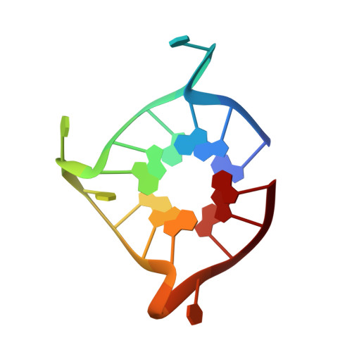
6PNK

Crystal structure of the G-quadruplex formed by (GGGTT)3GGG in complex with N-methylmesoporphryin IX

  • Classification: DNA
  • Organism(s): Tetrahymena thermophila
  • Mutation(s): No 

  • Deposited: 2019-07-02 Released: 2020-07-08 
  • Deposition Author(s): Yatsunyk, L.A., Lin, L.Y.
  • Funding Organization(s): National Institutes of Health/National Center for Research Resources (NIH/NCRR)

Experimental Data Snapshot

  • Method: X-RAY DIFFRACTION
  • Resolution: 2.39 Å
  • R-Value Free: 0.249 
  • R-Value Work: 0.235 
  • R-Value Observed: 0.236 

wwPDB Validation   3D Report Full Report


Ligand Structure Quality Assessment 


This is version 1.2 of the entry. See complete history


Literature

Biophysical and X-ray structural studies of the (GGGTT)3GGG G-quadruplex in complex with N-methyl mesoporphyrin IX.

Lin, L.Y.McCarthy, S.Powell, B.M.Manurung, Y.Xiang, I.M.Dean, W.L.Chaires, B.Yatsunyk, L.A.

(2020) PLoS One 15: e0241513-e0241513

  • DOI: https://doi.org/10.1371/journal.pone.0241513
  • Primary Citation of Related Structures:  
    6P45, 6PNK

  • PubMed Abstract: 

    The G-quadruplex (GQ) is a well-studied non-canonical DNA structure formed by G-rich sequences found at telomeres and gene promoters. Biological studies suggest that GQs may play roles in regulating gene expression, DNA replication, and DNA repair. Small molecule ligands were shown to alter GQ structure and stability and thereby serve as novel therapies, particularly against cancer. In this work, we investigate the interaction of a G-rich sequence, 5'-GGGTTGGGTTGGGTTGGG-3' (T1), with a water-soluble porphyrin, N-methyl mesoporphyrin IX (NMM) via biophysical and X-ray crystallographic studies. UV-vis and fluorescence titrations, as well as a Job plot, revealed a 1:1 binding stoichiometry with an impressively tight binding constant of 30-50 μM-1 and ΔG298 of -10.3 kcal/mol. Eight extended variants of T1 (named T2 -T9) were fully characterized and T7 was identified as a suitable candidate for crystallographic studies. We solved the crystal structures of the T1- and T7-NMM complexes at 2.39 and 2.34 Å resolution, respectively. Both complexes form a 5'-5' dimer of parallel GQs capped by NMM at the 3' G-quartet, supporting the 1:1 binding stoichiometry. Our work provides invaluable details about GQ-ligand binding interactions and informs the design of novel anticancer drugs that selectively recognize specific GQs and modulate their stability for therapeutic purposes.


  • Organizational Affiliation

    Department of Chemistry and Biochemistry, Swarthmore College, Swarthmore, Pennsylvania, United States of America.


Macromolecules

Find similar nucleic acids by:  Sequence   |   3D Structure  

Entity ID: 1
MoleculeChains LengthOrganismImage
DNA (5'-D(P*GP*GP*GP*TP*TP*GP*GP*GP*TP*TP*GP*GP*GP*TP*TP*GP*GP*G)-3')
A, B
18Tetrahymena thermophila
Sequence Annotations
Expand
  • Reference Sequence
Small Molecules
Ligands 4 Unique
IDChains Name / Formula / InChI Key2D Diagram3D Interactions
MMP (Subject of Investigation/LOI)
Query on MMP

Download Ideal Coordinates CCD File 
F [auth A],
J [auth B]
N-METHYLMESOPORPHYRIN
C35 H40 N4 O4
YNWHQWMCLCANDI-YIYRCNGCSA-N
FMT
Query on FMT

Download Ideal Coordinates CCD File 
K [auth B]FORMIC ACID
C H2 O2
BDAGIHXWWSANSR-UHFFFAOYSA-N
K
Query on K

Download Ideal Coordinates CCD File 
C [auth A]
D [auth A]
E [auth A]
G [auth B]
H [auth B]
C [auth A],
D [auth A],
E [auth A],
G [auth B],
H [auth B],
I [auth B]
POTASSIUM ION
K
NPYPAHLBTDXSSS-UHFFFAOYSA-N
NA
Query on NA

Download Ideal Coordinates CCD File 
L [auth B]SODIUM ION
Na
FKNQFGJONOIPTF-UHFFFAOYSA-N
Experimental Data & Validation

Experimental Data

  • Method: X-RAY DIFFRACTION
  • Resolution: 2.39 Å
  • R-Value Free: 0.249 
  • R-Value Work: 0.235 
  • R-Value Observed: 0.236 
  • Space Group: H 3 2
Unit Cell:
Length ( Å )Angle ( ˚ )
a = 60.93α = 90
b = 60.93β = 90
c = 194.342γ = 120
Software Package:
Software NamePurpose
PHENIXrefinement
XDSdata reduction
XDSdata scaling
PHASERphasing
Cootmodel building

Structure Validation

View Full Validation Report


Ligand Structure Quality Assessment 


Entry History & Funding Information

Deposition Data


Funding OrganizationLocationGrant Number
National Institutes of Health/National Center for Research Resources (NIH/NCRR)United States1R15CA208676-01A1

Revision History  (Full details and data files)

  • Version 1.0: 2020-07-08
    Type: Initial release
  • Version 1.1: 2020-11-25
    Changes: Database references, Derived calculations
  • Version 1.2: 2023-10-11
    Changes: Data collection, Database references, Refinement description