6OM8

Caenorhabditis Elegans UDP-Glucose Dehydrogenase in complex with UDP-Xylose


Experimental Data Snapshot

  • Method: X-RAY DIFFRACTION
  • Resolution: 2.45 Å
  • R-Value Free: 0.220 
  • R-Value Work: 0.194 
  • R-Value Observed: 0.194 

Starting Model: experimental
View more details

wwPDB Validation   3D Report Full Report


Ligand Structure Quality Assessment 


This is version 1.3 of the entry. See complete history


Literature

Conservation of Atypical Allostery inC. elegansUDP-Glucose Dehydrogenase.

Beattie, N.R.Keul, N.D.Hicks Sirmans, T.N.McDonald, W.E.Talmadge, T.M.Taujale, R.Kannan, N.Wood, Z.A.

(2019) ACS Omega 4: 16318-16329

  • DOI: https://doi.org/10.1021/acsomega.9b01565
  • Primary Citation of Related Structures:  
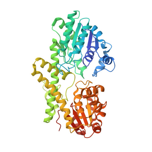
    6OM8

  • PubMed Abstract: 

    Human UDP-glucose dehydrogenase (hUGDH) oxidizes uridine diphosphate (UDP)-glucose to UDP-glucuronic acid, an essential substrate in the phase II metabolism of drugs. The activity of hUGDH is controlled by an atypical allosteric mechanism in which the feedback inhibitor UDP-xylose competes with the substrate for the active site and triggers a buried allosteric switch to produce an inactive complex (E Ω ). Previous comparisons with a nonallosteric UGDH identified six large-to-small substitutions that produce packing defects in the protein core and provide the conformational flexibility necessary for the allosteric transition. Here, we test the hypothesis that these large-to-small substitutions form a motif that can be used to identify allosteric UGDHs. Caenorhabditis elegans UGDH (cUGDH) conserves this motif with the exception of an Ala-to-Pro substitution in position 109. The crystal structures of unliganded and UDP-xylose bound cUGDH show that the A109P substitution is accommodated by an Asn-to-Ser substitution at position 290. Steady-state analysis and sedimentation velocity studies show that the allosteric transition is conserved in cUGDH. The enzyme also exhibits hysteresis in progress curves and negative cooperativity with respect to NAD + binding. Both of these phenomena are conserved in the human enzyme, which is strong evidence that these represent fundamental features of atypical allostery in UGDH. A phylogenetic analysis of UGDH shows that the atypical allostery motif is ancient and identifies a potential transition point in the evolution of the UGDH family.


  • Organizational Affiliation

    Department of Biochemistry & Molecular Biology and Institute of Bioinformatics, University of Georgia, Athens, Georgia 30602, United States.


Macromolecules
Find similar proteins by:  (by identity cutoff)  |  3D Structure
Entity ID: 1
MoleculeChains Sequence LengthOrganismDetailsImage
UDP-glucose 6-dehydrogenase
A, B, C, D, E
A, B, C, D, E, F, G, H, I, J, K, L
481Caenorhabditis elegansMutation(s): 0 
Gene Names: sqv-4F29F11.1
EC: 1.1.1.22
UniProt
Find proteins for Q19905 (Caenorhabditis elegans)
Explore Q19905 
Go to UniProtKB:  Q19905
Entity Groups  
Sequence Clusters30% Identity50% Identity70% Identity90% Identity95% Identity100% Identity
UniProt GroupQ19905
Sequence Annotations
Expand
  • Reference Sequence
Small Molecules
Ligands 1 Unique
IDChains Name / Formula / InChI Key2D Diagram3D Interactions
UDX
Query on UDX

Download Ideal Coordinates CCD File 
AA [auth H]
BA [auth H]
CA [auth I]
DA [auth I]
EA [auth J]
AA [auth H],
BA [auth H],
CA [auth I],
DA [auth I],
EA [auth J],
FA [auth J],
GA [auth K],
HA [auth K],
IA [auth L],
JA [auth L],
M [auth A],
N [auth A],
O [auth B],
P [auth B],
Q [auth C],
R [auth C],
S [auth D],
T [auth D],
U [auth E],
V [auth E],
W [auth F],
X [auth F],
Y [auth G],
Z [auth G]
URIDINE-5'-DIPHOSPHATE-XYLOPYRANOSE
C14 H22 N2 O16 P2
DQQDLYVHOTZLOR-OCIMBMBZSA-N
Experimental Data & Validation

Experimental Data

  • Method: X-RAY DIFFRACTION
  • Resolution: 2.45 Å
  • R-Value Free: 0.220 
  • R-Value Work: 0.194 
  • R-Value Observed: 0.194 
  • Space Group: P 2 21 21
Unit Cell:
Length ( Å )Angle ( ˚ )
a = 157.71α = 90
b = 168.17β = 90
c = 279.56γ = 90
Software Package:
Software NamePurpose
PHENIXrefinement
XDSdata scaling
Cootmodel building
PHENIXphasing
XDSdata reduction

Structure Validation

View Full Validation Report


Ligand Structure Quality Assessment 


Entry History & Funding Information

Deposition Data


Funding OrganizationLocationGrant Number
National Institutes of Health/National Institute of General Medical Sciences (NIH/NIGMS)United StatesR01GM11429

Revision History  (Full details and data files)

  • Version 1.0: 2019-05-22
    Type: Initial release
  • Version 1.1: 2019-10-30
    Changes: Data collection, Database references
  • Version 1.2: 2020-01-01
    Changes: Author supporting evidence
  • Version 1.3: 2023-10-11
    Changes: Data collection, Database references, Refinement description