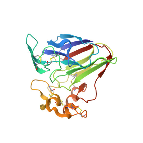
6O8A

Thaumatin native-SAD structure determined at 5 keV from microcrystals


Experimental Data Snapshot

  • Method: X-RAY DIFFRACTION
  • Resolution: 2.60 Å
  • R-Value Free: 0.216 
  • R-Value Work: 0.186 
  • R-Value Observed: 0.188 

wwPDB Validation   3D Report Full Report


This is version 1.2 of the entry. See complete history


Literature

Synchrotron microcrystal native-SAD phasing at a low energy.

Guo, G.Zhu, P.Fuchs, M.R.Shi, W.Andi, B.Gao, Y.Hendrickson, W.A.McSweeney, S.Liu, Q.

(2019) IUCrJ 6: 532-542

  • DOI: https://doi.org/10.1107/S2052252519004536
  • Primary Citation of Related Structures:  
    6O8A

  • PubMed Abstract: 

    De novo structural evaluation of native biomolecules from single-wavelength anomalous diffraction (SAD) is a challenge because of the weakness of the anomalous scattering. The anomalous scattering from relevant native elements - primarily sulfur in proteins and phospho-rus in nucleic acids - increases as the X-ray energy decreases toward their K -edge transitions. Thus, measurements at a lowered X-ray energy are promising for making native SAD routine and robust. For microcrystals with sizes less than 10 µm, native-SAD phasing at synchrotron microdiffraction beamlines is even more challenging because of difficulties in sample manipulation, diffraction data collection and data analysis. Native-SAD analysis from microcrystals by using X-ray free-electron lasers has been demonstrated but has required use of thousands of thousands of microcrystals to achieve the necessary accuracy. Here it is shown that by exploitation of anomalous microdiffraction signals obtained at 5 keV, by the use of polyimide wellmounts, and by an iterative crystal and frame-rejection method, microcrystal native-SAD phasing is possible from as few as about 1 200 crystals. Our results show the utility of low-energy native-SAD phasing with microcrystals at synchrotron microdiffraction beamlines.


  • Organizational Affiliation

    Biology Department, Brookhaven National Laboratory, Upton, NY 11973, USA.


Macromolecules
Find similar proteins by:  (by identity cutoff)  |  3D Structure
Entity ID: 1
MoleculeChains Sequence LengthOrganismDetailsImage
Thaumatin-1207Thaumatococcus danielliiMutation(s): 0 
UniProt
Find proteins for P02883 (Thaumatococcus daniellii)
Explore P02883 
Go to UniProtKB:  P02883
Entity Groups  
Sequence Clusters30% Identity50% Identity70% Identity90% Identity95% Identity100% Identity
UniProt GroupP02883
Sequence Annotations
Expand
  • Reference Sequence
Small Molecules
Ligands 1 Unique
IDChains Name / Formula / InChI Key2D Diagram3D Interactions
TLA
Query on TLA

Download Ideal Coordinates CCD File 
B [auth A]L(+)-TARTARIC ACID
C4 H6 O6
FEWJPZIEWOKRBE-JCYAYHJZSA-N
Experimental Data & Validation

Experimental Data

  • Method: X-RAY DIFFRACTION
  • Resolution: 2.60 Å
  • R-Value Free: 0.216 
  • R-Value Work: 0.186 
  • R-Value Observed: 0.188 
  • Space Group: P 41 21 2
Unit Cell:
Length ( Å )Angle ( ˚ )
a = 57.698α = 90
b = 57.698β = 90
c = 150.445γ = 90
Software Package:
Software NamePurpose
PHENIXrefinement
DIALSdata reduction
Aimlessdata scaling
SHARPphasing

Structure Validation

View Full Validation Report


Entry History & Funding Information

Deposition Data


Funding OrganizationLocationGrant Number
National Institutes of Health/National Human Genome Research Institute (NIH/NHGRI)United StatesGM107462

Revision History  (Full details and data files)

  • Version 1.0: 2019-05-08
    Type: Initial release
  • Version 1.1: 2019-07-31
    Changes: Data collection, Database references
  • Version 1.2: 2019-12-18
    Changes: Author supporting evidence