6MZ2

CTX-M-14 Class A Beta-Lactamase in Complex with Avibactam at pH 7.9


Experimental Data Snapshot

  • Method: X-RAY DIFFRACTION
  • Resolution: 0.83 Å
  • R-Value Free: 0.127 
  • R-Value Work: 0.116 
  • R-Value Observed: 0.117 

wwPDB Validation   3D Report Full Report


Ligand Structure Quality Assessment 


This is version 2.5 of the entry. See complete history


Literature

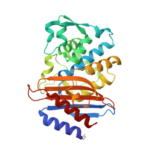
Mechanism of proton transfer in class A beta-lactamase catalysis and inhibition by avibactam.

Pemberton, O.A.Noor, R.E.Kumar M V, V.Sanishvili, R.Kemp, M.T.Kearns, F.L.Woodcock, H.L.Gelis, I.Chen, Y.

(2020) Proc Natl Acad Sci U S A 117: 5818-5825

  • DOI: https://doi.org/10.1073/pnas.1922203117
  • Primary Citation of Related Structures:  
    6MZ1, 6MZ2

  • PubMed Abstract: 

    Gram-negative bacteria expressing class A β-lactamases pose a serious health threat due to their ability to inactivate all β-lactam antibiotics. The acyl-enzyme intermediate is a central milestone in the hydrolysis reaction catalyzed by these enzymes. However, the protonation states of the catalytic residues in this complex have never been fully analyzed experimentally due to inherent difficulties. To help unravel the ambiguity surrounding class A β-lactamase catalysis, we have used ultrahigh-resolution X-ray crystallography and the recently approved β-lactamase inhibitor avibactam to trap the acyl-enzyme complex of class A β-lactamase CTX-M-14 at varying pHs. A 0.83-Å-resolution CTX-M-14 complex structure at pH 7.9 revealed a neutral state for both Lys73 and Glu166. Furthermore, the avibactam hydroxylamine- O -sulfonate group conformation varied according to pH, and this conformational switch appeared to correspond to a change in the Lys73 protonation state at low pH. In conjunction with computational analyses, our structures suggest that Lys73 has a perturbed acid dissociation constant (pK a ) compared with acyl-enzyme complexes with β-lactams, hindering its function to deprotonate Glu166 and the initiation of the deacylation reaction. Further NMR analysis demonstrated Lys73 pK a to be ∼5.2 to 5.6. Together with previous ultrahigh-resolution crystal structures, these findings enable us to follow the proton transfer process of the entire acylation reaction and reveal the critical role of Lys73. They also shed light on the stability and reversibility of the avibactam carbamoyl acyl-enzyme complex, highlighting the effect of substrate functional groups in influencing the protonation states of catalytic residues and subsequently the progression of the reaction.


  • Organizational Affiliation

    Department of Molecular Medicine, University of South Florida, Tampa, FL 33612.


Macromolecules
Find similar proteins by:  (by identity cutoff)  |  3D Structure
Entity ID: 1
MoleculeChains Sequence LengthOrganismDetailsImage
Beta-lactamase
A, B
263Escherichia coliMutation(s): 0 
Gene Names: blaCTX-M-14
EC: 3.5.2.6
UniProt
Find proteins for Q9L5C7 (Escherichia coli)
Explore Q9L5C7 
Go to UniProtKB:  Q9L5C7
Entity Groups  
Sequence Clusters30% Identity50% Identity70% Identity90% Identity95% Identity100% Identity
UniProt GroupQ9L5C7
Sequence Annotations
Expand
  • Reference Sequence
Small Molecules
Ligands 4 Unique
IDChains Name / Formula / InChI Key2D Diagram3D Interactions
NXL (Subject of Investigation/LOI)
Query on NXL

Download Ideal Coordinates CCD File 
C [auth A],
O [auth B]
(2S,5R)-1-formyl-5-[(sulfooxy)amino]piperidine-2-carboxamide
C7 H13 N3 O6 S
WJDGWXPPFHLLNL-RITPCOANSA-N
PO4
Query on PO4

Download Ideal Coordinates CCD File 
D [auth A],
P [auth B]
PHOSPHATE ION
O4 P
NBIIXXVUZAFLBC-UHFFFAOYSA-K
K
Query on K

Download Ideal Coordinates CCD File 
F [auth A]
G [auth A]
H [auth A]
I [auth A]
J [auth A]
F [auth A],
G [auth A],
H [auth A],
I [auth A],
J [auth A],
K [auth A],
L [auth A],
R [auth B],
S [auth B],
T [auth B],
U [auth B],
V [auth B],
W [auth B]
POTASSIUM ION
K
NPYPAHLBTDXSSS-UHFFFAOYSA-N
NA
Query on NA

Download Ideal Coordinates CCD File 
E [auth A],
M [auth A],
N [auth A],
Q [auth B]
SODIUM ION
Na
FKNQFGJONOIPTF-UHFFFAOYSA-N
Modified Residues  1 Unique
IDChains TypeFormula2D DiagramParent
PCA
Query on PCA
A, B
L-PEPTIDE LINKINGC5 H7 N O3GLN
Experimental Data & Validation

Experimental Data

  • Method: X-RAY DIFFRACTION
  • Resolution: 0.83 Å
  • R-Value Free: 0.127 
  • R-Value Work: 0.116 
  • R-Value Observed: 0.117 
  • Space Group: P 1 21 1
Unit Cell:
Length ( Å )Angle ( ˚ )
a = 45.161α = 90
b = 106.842β = 101.82
c = 47.913γ = 90
Software Package:
Software NamePurpose
HKL-2000data reduction
HKL-2000data scaling
PHASERphasing
PHENIXrefinement
PDB_EXTRACTdata extraction

Structure Validation

View Full Validation Report


Ligand Structure Quality Assessment 


Entry History & Funding Information

Deposition Data


Funding OrganizationLocationGrant Number
National Institutes of Health/National Institute Of Allergy and Infectious Diseases (NIH/NIAID)United StatesR01AI103158-04

Revision History  (Full details and data files)

  • Version 1.0: 2019-11-20
    Type: Initial release
  • Version 2.0: 2019-12-18
    Changes: Author supporting evidence, Polymer sequence
  • Version 2.1: 2019-12-25
    Changes: Database references
  • Version 2.2: 2020-03-11
    Changes: Database references, Structure summary
  • Version 2.3: 2020-03-18
    Changes: Database references
  • Version 2.4: 2020-04-01
    Changes: Database references
  • Version 2.5: 2023-10-11
    Changes: Data collection, Database references, Derived calculations, Refinement description