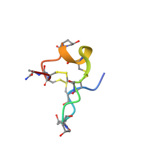
NMR Structure of mu-Conotoxin GIIIC: Leucine 18 Induces Local Repacking of the N-Terminus Resulting in Reduced NaVChannel Potency.
Harvey, P.J., Kurniawan, N.D., Finol-Urdaneta, R.K., McArthur, J.R., Van Lysebetten, D., Dash, T.S., Hill, J.M., Adams, D.J., Durek, T., Craik, D.J.(2018) Molecules 23
- PubMed: 30360356
- DOI: https://doi.org/10.3390/molecules23102715
- Primary Citation of Related Structures:
6MJD - PubMed Abstract:
μ-Conotoxins are potent and highly specific peptide blockers of voltage-gated sodium channels. In this study, the solution structure of μ-conotoxin GIIIC was determined using 2D NMR spectroscopy and simulated annealing calculations. Despite high sequence similarity, GIIIC adopts a three-dimensional structure that differs from the previously observed conformation of μ-conotoxins GIIIA and GIIIB due to the presence of a bulky, non-polar leucine residue at position 18. The side chain of L18 is oriented towards the core of the molecule and consequently the N-terminus is re-modeled and located closer to L18. The functional characterization of GIIIC defines it as a canonical μ-conotoxin that displays substantial selectivity towards skeletal muscle sodium channels (Na V ), albeit with ~2.5-fold lower potency than GIIIA. GIIIC exhibited a lower potency of inhibition of Na V 1.4 channels, but the same Na V selectivity profile when compared to GIIIA. These observations suggest that single amino acid differences that significantly affect the structure of the peptide do in fact alter its functional properties. Our work highlights the importance of structural factors, beyond the disulfide pattern and electrostatic interactions, in the understanding of the functional properties of bioactive peptides. The latter thus needs to be considered when designing analogues for further applications.
Organizational Affiliation:
Institute for Molecular Bioscience, The University of Queensland, Brisbane 4072, Australia. peta.harvey@imb.uq.edu.au.