6LEL

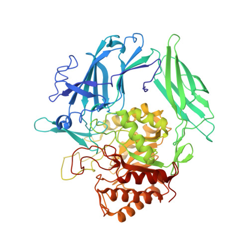
Structure of E. coli beta-glucuronidase complex with C6-hexyl uronic isofagomine


Experimental Data Snapshot

  • Method: X-RAY DIFFRACTION
  • Resolution: 2.50 Å
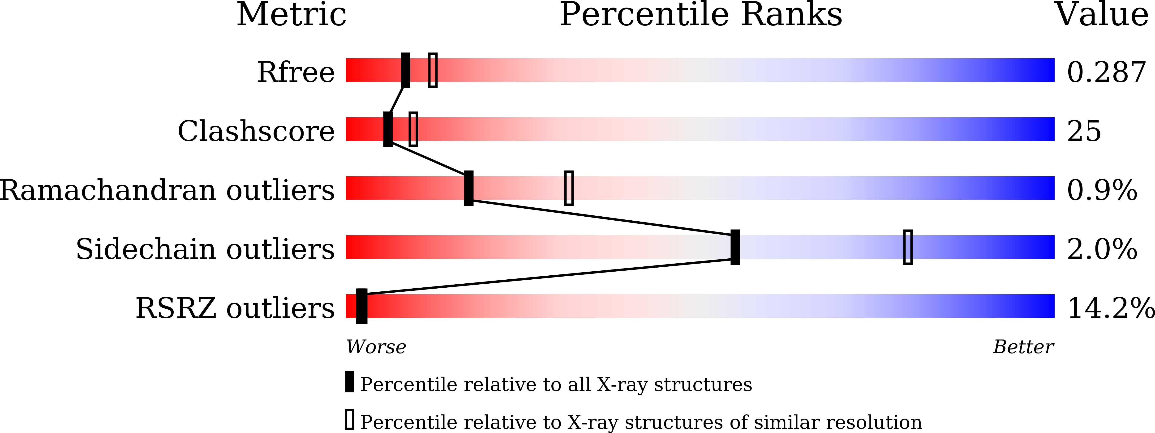
  • R-Value Free: 0.286 
  • R-Value Work: 0.238 
  • R-Value Observed: 0.240 

wwPDB Validation   3D Report Full Report


Ligand Structure Quality Assessment 


This is version 1.2 of the entry. See complete history


Literature

Entropy-driven binding of gut bacterial beta-glucuronidase inhibitors ameliorates irinotecan-induced toxicity.

Lin, H.Y.Chen, C.Y.Lin, T.C.Yeh, L.F.Hsieh, W.C.Gao, S.Burnouf, P.A.Chen, B.M.Hsieh, T.J.Dashnyam, P.Kuo, Y.H.Tu, Z.Roffler, S.R.Lin, C.H.

(2021) Commun Biol 4: 280-280

  • DOI: https://doi.org/10.1038/s42003-021-01815-w
  • Primary Citation of Related Structures:  
    6LD0, 6LD6, 6LDB, 6LDC, 6LDD, 6LEG, 6LEJ, 6LEL, 6LEM

  • PubMed Abstract: 

    Irinotecan inhibits cell proliferation and thus is used for the primary treatment of colorectal cancer. Metabolism of irinotecan involves incorporation of β-glucuronic acid to facilitate excretion. During transit of the glucuronidated product through the gastrointestinal tract, an induced upregulation of gut microbial β-glucuronidase (GUS) activity may cause severe diarrhea and thus force many patients to stop treatment. We herein report the development of uronic isofagomine (UIFG) derivatives that act as general, potent inhibitors of bacterial GUSs, especially those of Escherichia coli and Clostridium perfringens. The best inhibitor, C6-nonyl UIFG, is 23,300-fold more selective for E. coli GUS than for human GUS (K i  = 0.0045 and 105 μM, respectively). Structural evidence indicated that the loss of coordinated water molecules, with the consequent increase in entropy, contributes to the high affinity and selectivity for bacterial GUSs. The inhibitors also effectively reduced irinotecan-induced diarrhea in mice without damaging intestinal epithelial cells.


  • Organizational Affiliation

    Institute of Biological Chemistry, Academia Sinica, Taipei, Taiwan.


Macromolecules
Find similar proteins by:  (by identity cutoff)  |  3D Structure
Entity ID: 1
MoleculeChains Sequence LengthOrganismDetailsImage
Beta-D-glucuronidase
A, B
604Escherichia coliMutation(s): 0 
Gene Names: 
uidAA9R57_02750ACN68_26240ACN81_11215ACU57_22480AM270_03870AM464_25945AUQ13_03930BANRA_00079BANRA_00563BANRA_02251BB545_08625BET08_08370BHS87_09195BJJ90_13245BK292_18725BOH76_23450BON63_17270BON69_09035BON71_25965BUE81_02440BvCms2454_03287BvCmsHHP019_03955BvCmsKKP061_00687BvCmsKSP011_01150BvCmsKSP024_03693BvCmsKSP026_03508BvCmsKSP045_00685BvCmsKSP067_00118BvCmsNSP047_02620BvCmsSINP012_02859BvCmsSIP019_00145BVL39_19890BW690_15740BZL31_05290C4J69_24575C5N07_00485C7235_11830C9200_15785C9E25_02740C9Z12_02165CA593_19915CI694_12700COD46_02605CV83915_04776D2185_18760D3821_14300D3Y67_19800D9D20_10680D9I11_03210D9K48_06425DBQ99_12860DNQ41_12380DXT69_06115DXT71_07500E2119_03270E5P28_03750E5S47_08220EAI46_18055EAI52_05705EC3234A_33c01180EC3426_02616EEP23_13620EL75_2040EL79_2080EL80_2107ELT20_01525EPS71_15340ERS085365_00730ERS085416_00883ERS139211_01060ERS150873_00637EXX24_04480EXX78_14190EYD11_10940EYY78_00120FAX15_15905FV293_04650NCTC11181_05093NCTC13462_05935NCTC8500_02703NCTC9045_02849NCTC9062_00385NCTC9706_03872PU06_11555RG28_14030RK56_009440SAMEA3472044_02684SAMEA3472047_01270SAMEA3472080_01417SAMEA3484427_00776SAMEA3484429_00887SAMEA3752553_00640SAMEA3752559_02259SAMEA3753097_02320SK85_01835WR15_14365

EC: 3.2.1.31
UniProt
Find proteins for P05804 (Escherichia coli (strain K12))
Explore P05804 
Go to UniProtKB:  P05804
Entity Groups  
Sequence Clusters30% Identity50% Identity70% Identity90% Identity95% Identity100% Identity
UniProt GroupP05804
Sequence Annotations
Expand
  • Reference Sequence
Small Molecules
Ligands 1 Unique
IDChains Name / Formula / InChI Key2D Diagram3D Interactions
E8X (Subject of Investigation/LOI)
Query on E8X

Download Ideal Coordinates CCD File 
C [auth A],
D [auth B]
(2~{S},3~{S},4~{R},5~{R})-2-hexyl-4,5-bis(oxidanyl)piperidine-3-carboxylic acid
C12 H23 N O4
RRWNAAJMVJWKAM-VLEAKVRGSA-N
Experimental Data & Validation

Experimental Data

  • Method: X-RAY DIFFRACTION
  • Resolution: 2.50 Å
  • R-Value Free: 0.286 
  • R-Value Work: 0.238 
  • R-Value Observed: 0.240 
  • Space Group: C 1 2 1
Unit Cell:
Length ( Å )Angle ( ˚ )
a = 165.781α = 90
b = 76.72β = 124.64
c = 124.888γ = 90
Software Package:
Software NamePurpose
HKL-2000data scaling
PHENIXrefinement
PDB_EXTRACTdata extraction
Blu-Icedata collection
PHENIXphasing

Structure Validation

View Full Validation Report


Ligand Structure Quality Assessment 


Entry History & Funding Information

Deposition Data


Funding OrganizationLocationGrant Number
Ministry of Science and Technology (Taiwan)Taiwan108-2113-M-001-001

Revision History  (Full details and data files)

  • Version 1.0: 2021-01-27
    Type: Initial release
  • Version 1.1: 2021-08-11
    Changes: Database references
  • Version 1.2: 2023-11-22
    Changes: Data collection, Refinement description