6L88

Crystal structure of mineralocorticoid receptor ligand binding domain in complex with esaxerenone


Experimental Data Snapshot

  • Method: X-RAY DIFFRACTION
  • Resolution: 3.00 Å
  • R-Value Free: 0.307 
  • R-Value Work: 0.269 
  • R-Value Observed: 0.271 

wwPDB Validation   3D Report Full Report


Ligand Structure Quality Assessment 


This is version 1.2 of the entry. See complete history


Literature

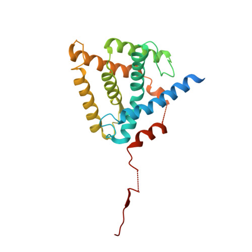
Crystal structure of the mineralocorticoid receptor ligand-binding domain in complex with a potent and selective nonsteroidal blocker, esaxerenone (CS-3150).

Takahashi, M.Ubukata, O.Homma, T.Asoh, Y.Honzumi, M.Hayashi, N.Saito, K.Tsuruoka, H.Aoki, K.Hanzawa, H.

(2020) FEBS Lett 594: 1615-1623

  • DOI: https://doi.org/10.1002/1873-3468.13746
  • Primary Citation of Related Structures:  
    6L88

  • PubMed Abstract: 

    Activation of the mineralocorticoid receptor (MR) has long been considered a risk factor for cardiovascular diseases. It has been reported that the novel MR blocker esaxerenone shows high potency and selectivity for MR in vitro as well as great antihypertensive and renoprotective effects in salt-sensitive hypertensive rats. Here, we determined the cocrystal structure of the MR ligand-binding domain (MR-LBD) with esaxerenone and found that esaxerenone binds to MR-LBD in a unique manner with large side-chain rearrangements, distinct from those of previously published MR antagonists. This structure also displays an antagonist form that has not been observed for MR previously. Such a unique binding mode of esaxerenone provides great insight into the novelty, potency, and selectivity of this novel antihypertensive drug.


  • Organizational Affiliation

    Structure-Based Drug Design Group, Organic Synthesis Department, Daiichi Sankyo RD Novare Co., Ltd., Tokyo, Japan.


Macromolecules
Find similar proteins by:  (by identity cutoff)  |  3D Structure
Entity ID: 1
MoleculeChains Sequence LengthOrganismDetailsImage
Mineralocorticoid receptor
A, B, C, D
251Homo sapiensMutation(s): 2 
Gene Names: NR3C2MCRMLR
UniProt & NIH Common Fund Data Resources
Find proteins for P08235 (Homo sapiens)
Explore P08235 
Go to UniProtKB:  P08235
PHAROS:  P08235
GTEx:  ENSG00000151623 
Entity Groups  
Sequence Clusters30% Identity50% Identity70% Identity90% Identity95% Identity100% Identity
UniProt GroupP08235
Sequence Annotations
Expand
  • Reference Sequence
Small Molecules
Binding Affinity Annotations 
IDSourceBinding Affinity
E6R BindingDB:  6L88 IC50: min: 2.4, max: 1000 (nM) from 3 assay(s)
Experimental Data & Validation

Experimental Data

  • Method: X-RAY DIFFRACTION
  • Resolution: 3.00 Å
  • R-Value Free: 0.307 
  • R-Value Work: 0.269 
  • R-Value Observed: 0.271 
  • Space Group: P 21 3
Unit Cell:
Length ( Å )Angle ( ˚ )
a = 160.888α = 90
b = 160.888β = 90
c = 160.888γ = 90
Software Package:
Software NamePurpose
REFMACrefinement
XSCALEdata scaling
PDB_EXTRACTdata extraction
XDSdata reduction
PHASERphasing

Structure Validation

View Full Validation Report


Ligand Structure Quality Assessment 


Entry History 

Deposition Data

Revision History  (Full details and data files)

  • Version 1.0: 2020-02-12
    Type: Initial release
  • Version 1.1: 2020-06-10
    Changes: Database references
  • Version 1.2: 2023-11-22
    Changes: Data collection, Database references, Refinement description