


0.98 A three-dimensional structure of horse heart cytochrome C at 110K
Sugawara, Y., Endo, S.To be published.
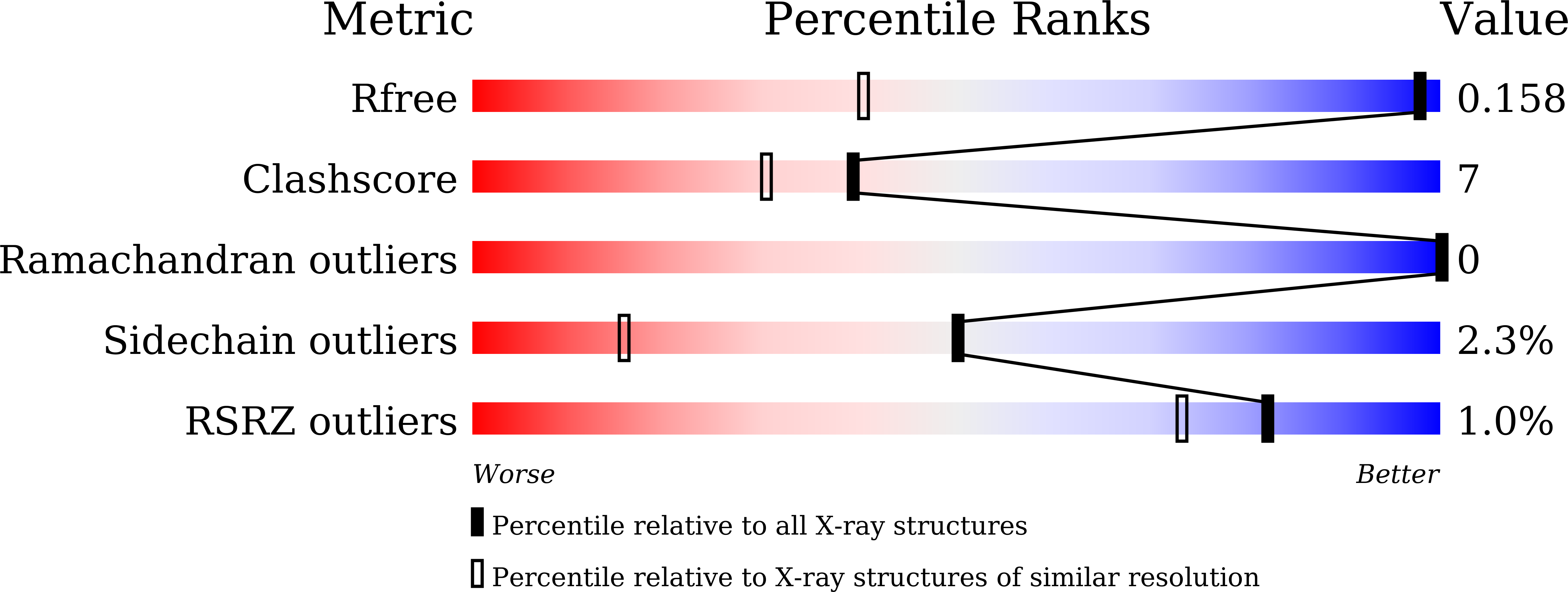
Experimental Data Snapshot
Entity ID: 1 | |||||
|---|---|---|---|---|---|
| Molecule | Chains | Sequence Length | Organism | Details | Image |
| Cytochrome c | 105 | Equus caballus | Mutation(s): 1 | 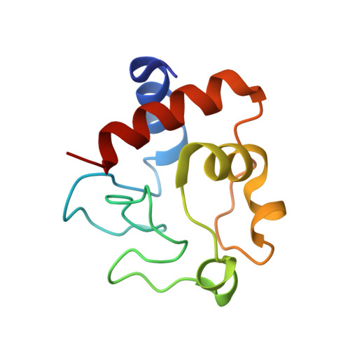 | |
UniProt | |||||
Find proteins for P00004 (Equus caballus) Explore P00004 Go to UniProtKB: P00004 | |||||
Entity Groups | |||||
| Sequence Clusters | 30% Identity50% Identity70% Identity90% Identity95% Identity100% Identity | ||||
| UniProt Group | P00004 | ||||
Sequence AnnotationsExpand | |||||
| |||||
| Ligands 1 Unique | |||||
|---|---|---|---|---|---|
| ID | Chains | Name / Formula / InChI Key | 2D Diagram | 3D Interactions | |
| HEC Query on HEC | B [auth A] | HEME C C34 H34 Fe N4 O4 HXQIYSLZKNYNMH-LJNAALQVSA-N |  | ||
| Length ( Å ) | Angle ( ˚ ) |
|---|---|
| a = 56.887 | α = 90 |
| b = 56.887 | β = 90 |
| c = 42.487 | γ = 90 |
| Software Name | Purpose |
|---|---|
| SHELXL | refinement |
| CrysalisPro | data reduction |
| CrysalisPro | data scaling |
| MOLREP | phasing |
RCSB PDB (citation) is hosted by
RCSB PDB is a member of the

